
Tissue integration design for seamless implant fixation

a meshtissue and implantable device technology, applied in the direction of prosthesis, shoulder joints, osteosynthesis devices, etc., can solve the problems of implant failure, compromise of function, loosening of implants,

Active Publication Date: 2016-06-14
BIEDERMANN TECH GMBH & CO KG
View PDF58 Cites 11 Cited by
  • Summary
  • Abstract
  • Description
  • Claims
  • Application Information

AI Technical Summary

Benefits of technology

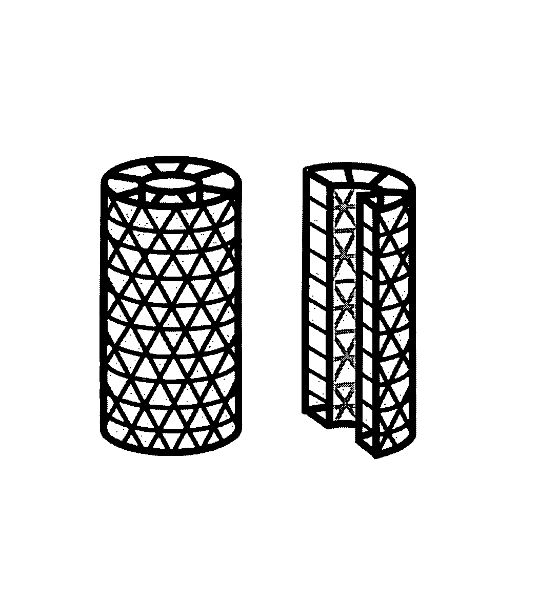
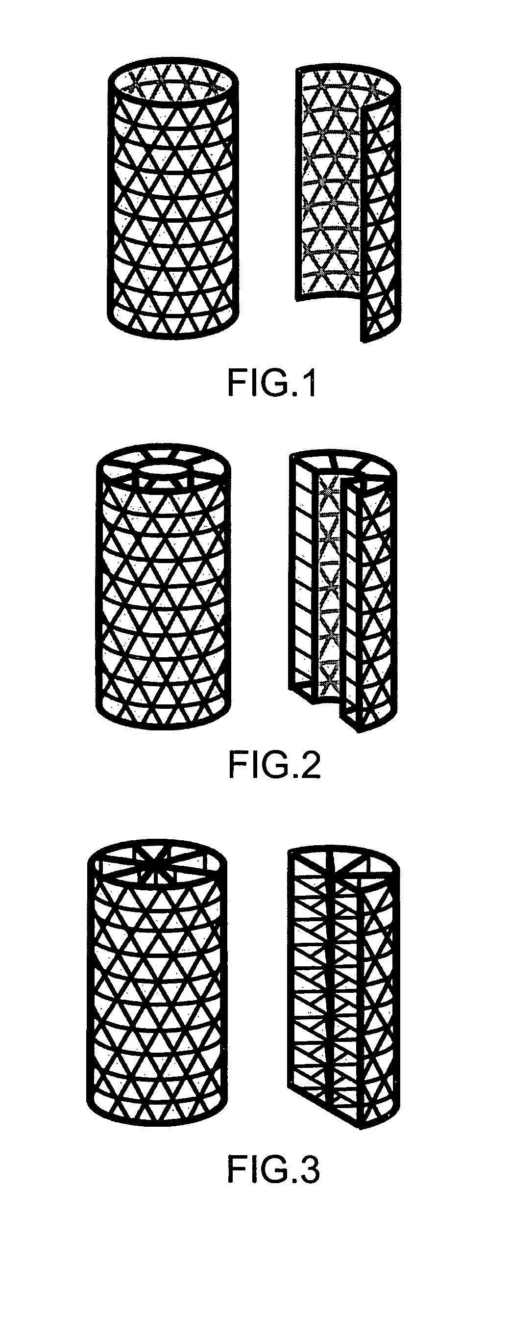
The present invention is about an improved muscle-skeletal implant that can adapt to the mechanical demands of a person's life. The implant has a fenestrated shell component that allows for the integration of the implant fixation with the host tissue. It can be made of titanium, ceramic material, or cobalt-chromium, and can have a honeycomb pattern or any other pattern. The implant can also have bone tissue or synthetic materials in the biologic core. It can be used for dental or joint replacement implants. The implant can have bone implant hardware, such as bone nails, screws, rods, or plates. It can also have a coating of material on its surface, such as hydroxyapatite. The implant can have one or more two-dimensional or three-dimensional mechanical structures that maintain their form against their own weight.

Problems solved by technology

The inability to achieve and maintain fixation results in a number of complications which include pain, implant loosening, implant failure, and compromise of function.
The major problem inherent in all implant devices is the gradual loosening over time.
This problem is especially prevalent where the implant is subjected to large functional loads and sheer stresses.
Cranio-facial implants are particularly prone to this problem.
Other orthopedic implants such as plates, screws, nails, pins, etc., are also subjected to fixation problems.
Both these conventional methods are usually characterized by having the greatest degree of fixation immediately: after implantation, but suffer from loss of fixation in the months and years after implantation.
Additionally, there is a disadvantage with the use of cements such as methylmethacrylate due to its potential toxicity.
Their disadvantages are their generally weaker mechanical properties, their poorer biocompatibility, and their much shorter history of clinical use.
Despite the plethora of existing approaches for securing an implanted structure into bone, fixation failures commonly occur.
These failures are primarily due to implant loosing caused by the inability of the bone to withstand the physiological loads at the bone / implant interface.
One factor in such failures is that the new bone ingrowth surrounding the implant is limited to the surface.
The inadequate fixation with the host tissue has been one of the major limitations of these devices.
The principal components of inadequate fixation include: 1) inadequate bonding with host tissue; 2) non-optimal biomechanical properties; and 3) incompatible biologic properties.
With respect to bonding of host tissue, conventional implants have been limited to bonding to the surface of the implant only, with very limited or no tissue ingrowth.
Additionally, there is always an interface between the implant and the host tissue, and the interface is always biomechanically and biologically inferior to the implant and host tissue.
With respect to biomechanical properties, conventional implants are bulky and stiff in order to offset fatigue, which creates a disparity between the mechanical properties of the implant and the host tissue.
This disparity results in stress risers, stress shielding, and bone atrophy.
Finally, with respect to the biologic properties, conventional implants either do not support or poorly support tissue biology.
These conventional implants are space-occupying devices which alter local tissue biology, do not accommodate the quality of the host tissue, and do not remodel with the host tissue.
Additionally, they do not allow for the application of biological factors which could enhance implant function.
The result is a biological and biomechanical disparity between the implant and host tissue which culminates in loss of implant fixation.

Method used


Image

  • Tissue integration design for seamless implant fixation
  • Tissue integration design for seamless implant fixation
  • Tissue integration design for seamless implant fixation

Examples


Embodiment Construction

[0034]As used herein, “a” or “an” is defined herein as one or more.

[0035]As used herein, “core” is defined as the internal space within or in-between a shell component to contain, at least in part, a material that induces or promotes biological activity

[0036]As used herein, the term “fenestrated” is defined as the quality of possessing macroscopic perforations or holes in an otherwise solid, hollow, or mesh component. In reference to the outer and / or inner shell component of the present invention, “fenestrated” refers to the quality of possessing holes or perforations through which material can grow into or out of the inside of the shell.

[0037]As used herein, the term “fenestrated shell component”, means a component comprising one or more than one fenestrated shell.

[0038]As used herein, the term “implant” is defined broadly, encompassing any and all devices implanted into humans or animals. These include, but are not limited to, orthopaedic implants and dental implants.

[0039]As used...


PUM

PropertyMeasurementUnit
weightaaaaaaaaaa
ultra-high aaaaaaaaaa
fixation stabilityaaaaaaaaaa

Abstract

The present invention relates to orthopaedic implants having a fenestrated hollow shell and a biologic core. These design features provide an improved interface between the implant and the surrounding tissue, aiding fixation, and provide a vehicle for applying new bone healing and enhancing modalities, such as gene therapy, tissue engineering, and growth factors.

Description

CROSS-REFERENCE TO RELATED APPLICATIONS[0001]This application claims priority to U.S. provisional application Ser. No. 60 / 482,414, filed Jun. 25, 2003.TECHNICAL FIELD[0002]The present invention relates to the development and use of a mesh-tissue integration implantable device. The implant has the advantages of providing superior fixation and enhanced biological and biomechanical function which increase its longevity relative to conventional implants.BACKGROUND OF THE INVENTION[0003]Medical and dental implants are widely used today. Typical examples would be the implants used for joint replacements, fractures fixation, and bone reconstruction. Joint replacement implants typically comprise two parts: a metal or ceramic part formed with an articulating surface designed to be received in and rub against a complementary load-bearing plastic surface of either an all-plastic part or a metal part with a plastic surface. In the case of metal implants, the type of metal may include titanium a...

Claims


Application Information

Patent Timeline
Patent Type & Authority Patents(United States)
IPC IPC(8): A61F2/44A61B17/68A61F2/36A61F2/30A61B17/70A61F2/28A61F2/42A61F2/40A61F2/38A61F2/32A61B17/72A61B17/74A61B17/80A61B17/86A61FA61F2/00
CPCA61B17/68A61F2/30907A61B17/72A61B17/7233A61B17/7291A61B17/742A61B17/80A61B17/866A61F2/28A61F2/30771A61F2/32A61F2/38A61F2/40A61F2/42A61F2/44A61F2002/2835A61F2002/30677A61F2002/30678A61F2002/30784A61F2002/3085A61F2310/00179A61F2310/00353A61F2310/00407A61F2310/00413A61F2310/00592A61F2310/00796A61F2310/00359A61F2/2846A61F2002/3024A61F2002/3092A61F2002/30235
Inventor LINDSEY, RONALD W.GUGALA, ZBIGNIEWLATTA, LOREN L.
Owner BIEDERMANN TECH GMBH & CO KG
Learn More