In a DR screening
system, for example, an image is deemed as inadequate when it is difficult or impossible to make a reliable clinical judgment regarding the presence or absence of DR in the image.
Major causes of inadequate image quality in
retinal screening images include illumination crescents due to small
pupil size, loss of contrast due to poor focus or movement of the eye or media
opacity, and imaging of part of the
eyelid and
eyelash due to blinking, as well as insufficient illumination.
Inadequate images reduce the utility of automatic screening systems because they have to be discarded from analysis, or cause preventable errors in said systems.
Further, image quality is also negatively correlated to subject's age, and presence and level of
retinal disease.
Taking more images after
visual inspection of image quality may solve some of the problems but at the expense of time since more images are needed, the comfort of the patient since more flashes of
high intensity light are needed, and, paradoxically, detriment of image quality since the patient's
pupil may not reach optimum natural dilation after just a three or four images are taken.
Therefore, visual aids currently available in desktop non-mydriatic cameras are insufficient to ensure efficient imaging time and quality.
The likelihood of high percentages of unusable images prevent wide adoption of retinal screening at the primary
care setting and limit the clinical utility of currently available systems.
Today's commercial cameras have limited clinical utility at the primary
care setting because they do not ensure that rates of unusable images will be low enough to justify the investment of $25,000 or more per camera.
Even low-cost camera alternatives are difficult to justify when their efficiency depends heavily on photographer skills.
Often the photographer taking the image will not appreciate the requisite criteria for image quality required by the
end user, whether an ophthalmologist or a highly trained grader or reader of the retinal images.
What may appear acceptable to the photographer may be deemed unacceptable or entirely unusable by the grader.
In telemedicine, transmitting an unacceptable quality image may mean, at worse a
missed diagnosis, or at best the need to retake the image or images at an inconvenience to a patient who will have to return to the clinic for re-imaging.
In longitudinal studies where every image is critically important, losing an image for a given examination period may result in loss of that individual from the study or a reduction in the statistical power of the results.
2) grader- and photographer-dependent issues comprise
visual perception, training, and experience;
4) patient-depended issues comprise lens and media opacities, ocular aberrations, and retinal pigmentation.
While certain types of technical errors, e.g. poor alignment,
pupil centering, blinks, can be corrected by re-acquiring the
retinal image, others such as camera artifacts, e.g. glares, dust, scratches on
optics, etc., cannot.
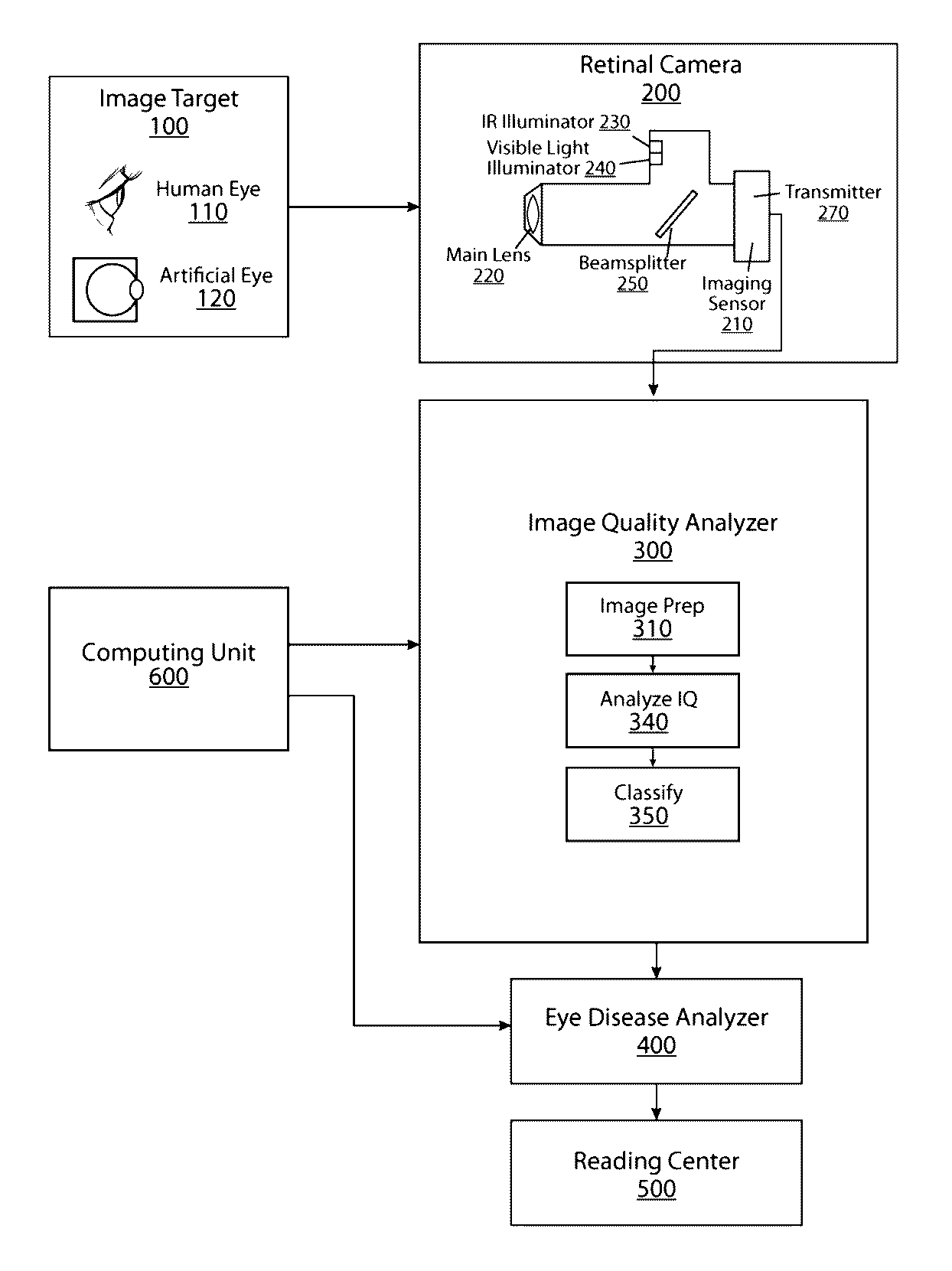
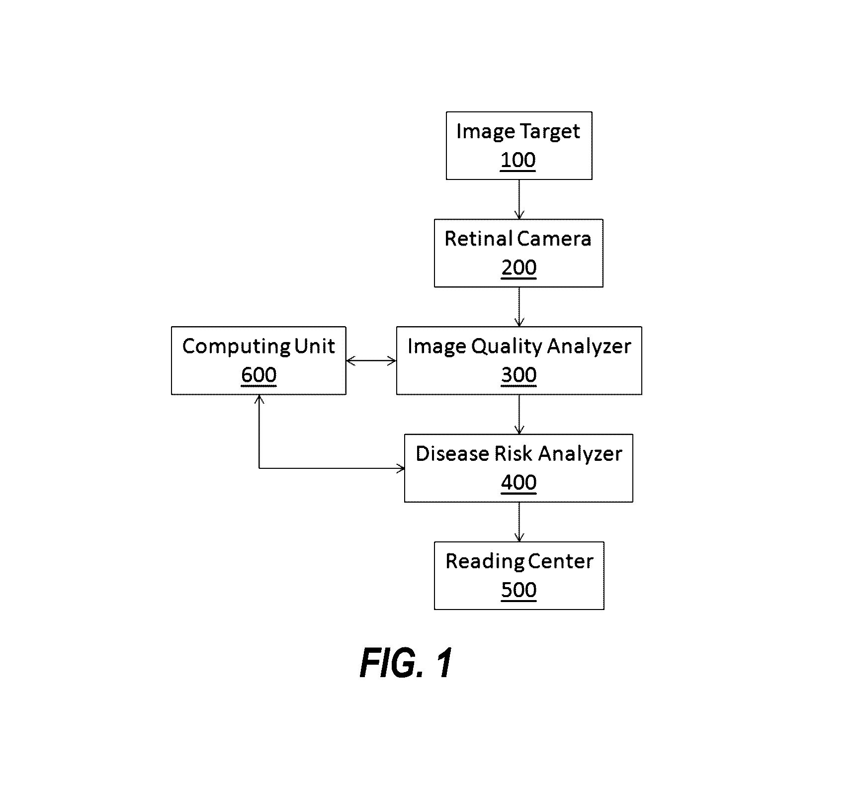
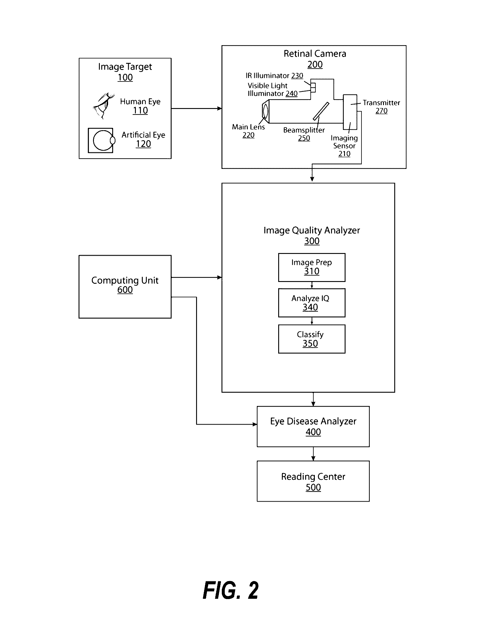
Currently, there are no retinal cameras that provide real time image
quality assessment during alignment or after an image is acquired.
Some desktop retinal cameras provide visual aids for alignment and focus but these are not sufficient to guide the photographer to ensure maximum image quality.
Depending on the application, the quality of an image is deemed deficient when it becomes difficult or impossible to make a meaningful clinical judgment regarding the presence or absence of signs of
pathology in the image, as listed in Table 1.
Reference image-based methods (i.e. a quality comparison with the optimal image of each
retina is made using various quantitative measures) are disadvantageous because a limited number of the good quality images might not be a “good” reference for the natural large variance encountered in retinal images acquired from screening.
The practical utility of these techniques is very low because they are computationally intensive (not suited for real-time feedback to a photographer) and have an inherent uncertainty regarding a valid or successful segmentation step (such as finding small vessels to determine the quality of a
retinal image).
The utility of these methods is limited to “recreational” photography where the image quality factors include image brightness, contrast, and focus.
These recreational photography methods fail to consider other factors involved in assessing image quality of medical images in general and retinal images in particular.
A
retinal image can have adequate levels of brightness, contrast, or focus but if one of these areas is not present, the image is unusable for practical purposes.
Further, said combination of image quality factors has not been reported in the literature or applied in practice.
However, this technique is computationally intensive and not suitable for real-time applications.
Though effective in evaluating image quality (0.997 area under the area under the
receiver operating curve, AUC), this approach was found to require up to 30 seconds to perform the necessary calculations, thus exceeding the practical limits for clinical, real-time applications.
This technique is computationally burdensome and must integrate explicitly all possible factors, each treated independently with a different
algorithm.
Detecting the failure of the segmentation in case of low image quality is not trivial and limits the robustness of the approach and its clinical utility.

