
Method for high-throughput protein detection with two antibody microarrays

a protein detection and antibody technology, applied in the field of detecting a plurality of ligands, can solve the problems of being unable to identify individual phosphorylated proteins, being extremely difficult to achieve, and only applicable, so as to improve the quality of protein detection assays

Active Publication Date: 2017-07-25
WANG YINGJIAN
View PDF2 Cites 0 Cited by
  • Summary
  • Abstract
  • Description
  • Claims
  • Application Information

AI Technical Summary

Benefits of technology

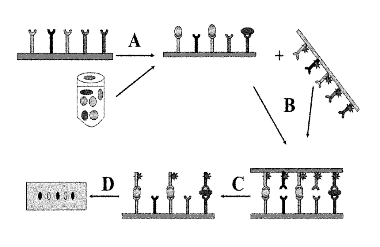
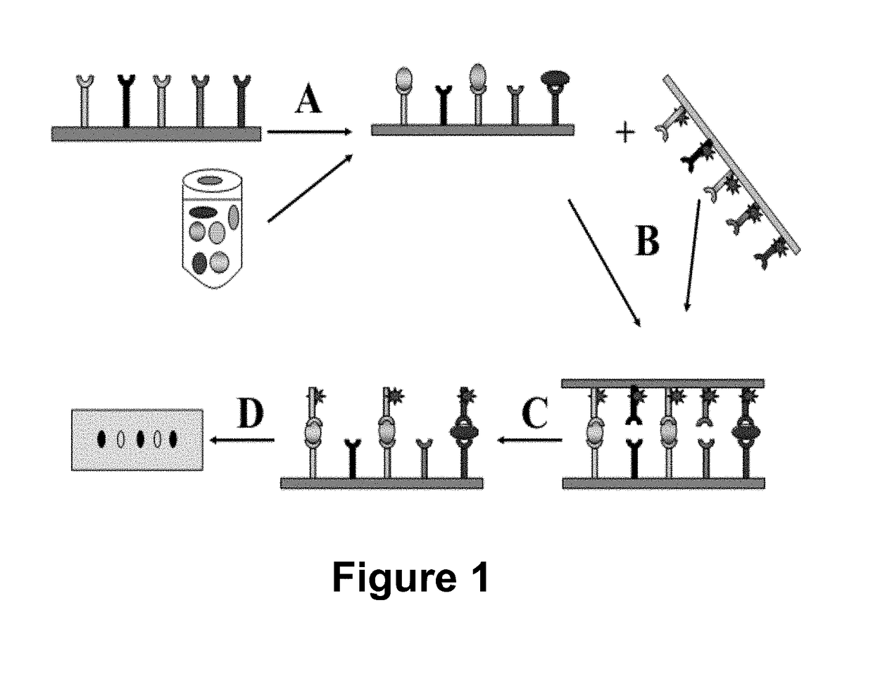
[0012]It is an object of the invention to provide methods that enable one to detect proteins and their properties, and more particularly, to detect and compare the presences of proteins in biological samples. The methods of the invention are designed to substantially improve the quality of protein detection assays. For example, two arrays of antibodies immobilized on two solid supports are used in the methods so that two antibodies are employed to detect one antigen while avoiding the interference from other antibodies on the arrays.

Problems solved by technology

The protein expression pattern of a cell determines its shape and function; and abnormal protein expressions cause cells to malfunction, resulting in diseases.
However, these methods are usually only applicable to the analysis of one or a few proteins at a time.
Although antibodies specific for phosphorylated amino acids, such as PY20 and 4G10, can reveal multiple phosphorylated proteins, they alone are unable to identify individual phosphorylated proteins.
However, this has turned out to be extremely difficult to achieve.
However, it requires complicated procedures and it is necessary to determine the identities of the proteins displayed on the two-dimensional gel, which is difficult to achieve for most proteins.
The most challenging problem of the current protein array technology is low specificity.
The problem is due primarily to the so-called “non-specific” binding of capture and detection reagents (e.g. antibodies).

Method used


Image

  • Method for high-throughput protein detection with two antibody microarrays
  • Method for high-throughput protein detection with two antibody microarrays
  • Method for high-throughput protein detection with two antibody microarrays

Examples


example 1

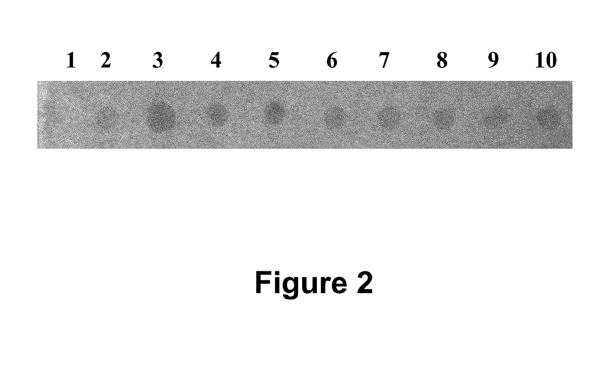
[0066]In this example (FIG. 2) a cell lysate prepared from 293T human cells was immobilized at 10 spots on a nitrocellulose membrane to form a ligand array. About 2 micrograms of protein lysate was immobilized at each spot. 10 different rabbit polyclonal antibodies against 10 different cellular proteins were immobilized on a nylon membrane to form a dissociable antibody array. After blocking, the dissociable antibody array was laid on top of the ligand array to make contact. After 60 minutes incubation during which time antibodies bound to their respective antigens, the dissociable antibody array was removed from the ligand array. The ligand-bound antibodies stayed on the ligand array. Any non-specific binding antibodies were washed off; and the rabbit antibodies were detected with HRP-conjugated goat-anti-rabbit secondary antibodies. In this example, TMB was used as HRP substrate.

example 2

[0067]In this example, unconjugated secondary antibodies were used to capture serum IgG while HRP-conjugated secondary antibodies were used as the detection / dissociable antibodies. Goat-anti-mouse and goat-anti-rabbit IgG(Fc) antibodies were spotted on a silane-coated glass slide and covalently immobilized on it to produce the capture antibody microarray. To make a dissociable antibody microarray, HRP-conjugated goat-anti-mouse or goat-anti-rabbit IgG [F(ab′)2] antibodies were spotted on a nylon membrane with a glass slide as backing. About 80 nl antibody solutions were deposited at each spot 2 mm apart; each antibody was spotted in duplicate. The capture and detection antibodies recognize different epitopes (Fc and F(ab′)2, respectively) of the IgG ligand. Mouse serum (1:1000 dilution) was used as target ligand. HRP substrate Tetramethylbenzidine Dihydro-chloride was used to visualize the bound HRP-conjugated detection antibody. As shown in FIG. 3A, signals were detected at the pos...

example 3

[0069]In this example, two antibody arrays were prepared and used to detect proteins according to the method disclosed here. Antibodies from commercial sources (about 0.2 μg / μl) were arrayed on membranes using a robotic arrayer to make both capture and dissociable antibody arrays. Nitrocelluose membrane was used as capture antibody array support; and nylon membrane was used as dissociable antibody array support. All antibodies on the capture antibody array are from rabbit; and all antibodies on the dissociable antibody array are from mouse. Decreasing amounts of antibodies were immobilized in each row on the capture antibody arrays. Rows 1 and 3 are antibodies against Connexin43 protein, and rows 2 and 4 are antibodies against GST protein. Same amount of antibodies (40 ng) were immobilized in each row on dissociable antibody array. Antibodies were immobilized by non-covalent bonds between nylon / nitrocelluose membranes and the antibodies. Antibody arrays were either used immediately ...


PUM

PropertyMeasurementUnit
thicknessaaaaaaaaaa
covalentaaaaaaaaaa
shapeaaaaaaaaaa

Abstract

The invention provides a method for detecting one or more biological ligands, where the method generally uses two arrays of biological reagents. The two arrays have two different functionalities: the first array captures the ligands on the array; and the second array delivers detecting reagents to the captured ligands. Use of arrays affords the method high-throughput detection capability; and the use of two different arrays ensures high specificity. The first array has a support structure fixed with a first set of reagents, each at a pre-determined position on the support. The second array has a support structure fixed with a second set of reagents, each at a pre-determined position on the support. The first array contains reagent(s) binding to a ligand, while the second array contains second reagent(s) binding to the same ligand. In preferred embodiments, antibodies are used as reagents, and used in detecting proteins in protein samples.

Description

FIELD OF THE INVENTION[0001]The present invention generally relates to methods for detecting a plurality of ligands and more particularly to methods for detecting the expressions, activities and functions of multiple proteins.BACKGROUND OF THE INVENTION[0002]The availability of a large number of biological reagents, such as hundreds of thousands of cloned DNA sequences, numerous antibodies and recombinant proteins, millions of compounds obtained through combinatory chemical synthesis, has promoted the development of technologies that make use of these reagents in biological research, clinical diagnostics and drug development. Special position-addressable arrays of biological reagents have been designed, in which each of the reagents is placed at a pre-defined position so that it can be identified later by the position. For example, in a DNA array, a large number of cDNA or oligos are immobilized, each at a pre-defined position and can be identified later by that position. DNA arrays...

Claims


Application Information

Patent Timeline
Patent Type & Authority Patents(United States)
IPC IPC(8): G01N33/53G01N33/545G01N33/68G01N33/543
CPCG01N33/6845G01N33/545G01N33/54353G01N2440/14G01N33/54306G01N33/54366
Inventor WANG, YINGJIAN
Owner WANG YINGJIAN
Learn More