Sparse deconvolution spatial light microscopy in two and three dimensions

a spatial light microscopy and deconvolution technology, applied in the field of three-dimensional imaging, can solve the problems of interference-based microscopy, fluorescence probes that are often harmful to living specimens, and difficult tomographic imaging of live cells

Active Publication Date: 2017-07-25
THE BOARD OF TRUSTEES OF THE UNIV OF ILLINOIS
View PDF1 Cites 2 Cited by
  • Summary
  • Abstract
  • Description
  • Claims
  • Application Information

AI Technical Summary

Problems solved by technology

More particularly, transparency has rendered tomographic imaging of live cells particularly challenging.
While confocal fluorescence imaging is a common approach to achieve sectioning, it requires fluorescence probes that are often harmful to a living specimen.
Interference-based microscopy is afflicted by optical degradation and noise, both introduced by the instrument.
However, due to the laser light illumination, the noise in the images limits the performance of the deconvolution and the overall resolution gain.
The method of Haldar (2010), however, fails to exploit the sparsity information of biological specimen because the quadratic regularization employed implicitly enforces smoothness on the unknown phase field, even though the phase field is typically not smooth at object boundaries such as fine-scale cell boundaries and intracellular structures.
Haldar (2010), moreover, uses a nonlinear conjugate gradient method to obtain both phase and magnitude, which generally contains several approximations and heuristics, and is therefore not as well characterized as linear methods in terms of stability and convergence.

Method used


Image

  • Sparse deconvolution spatial light microscopy in two and three dimensions
  • Sparse deconvolution spatial light microscopy in two and three dimensions
  • Sparse deconvolution spatial light microscopy in two and three dimensions

Examples


examples

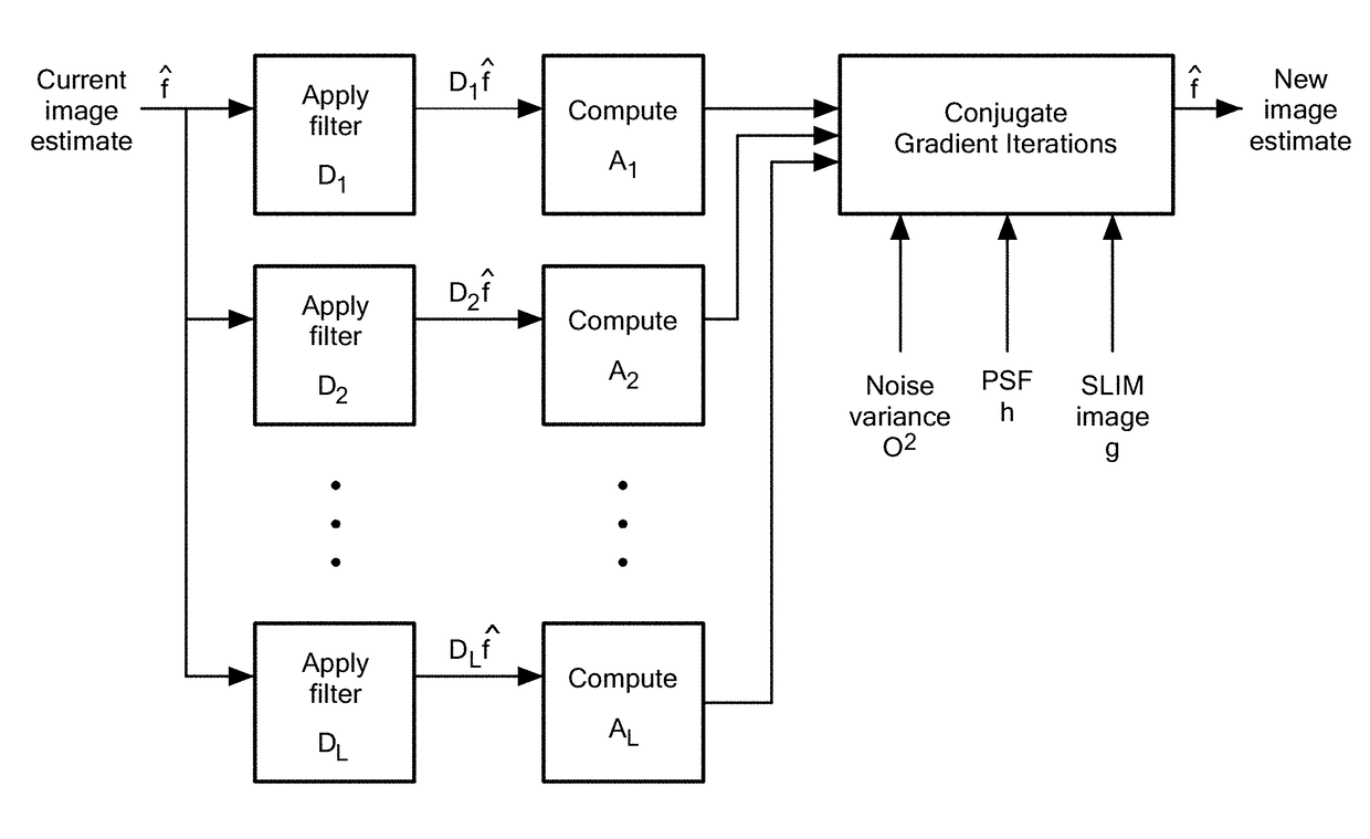
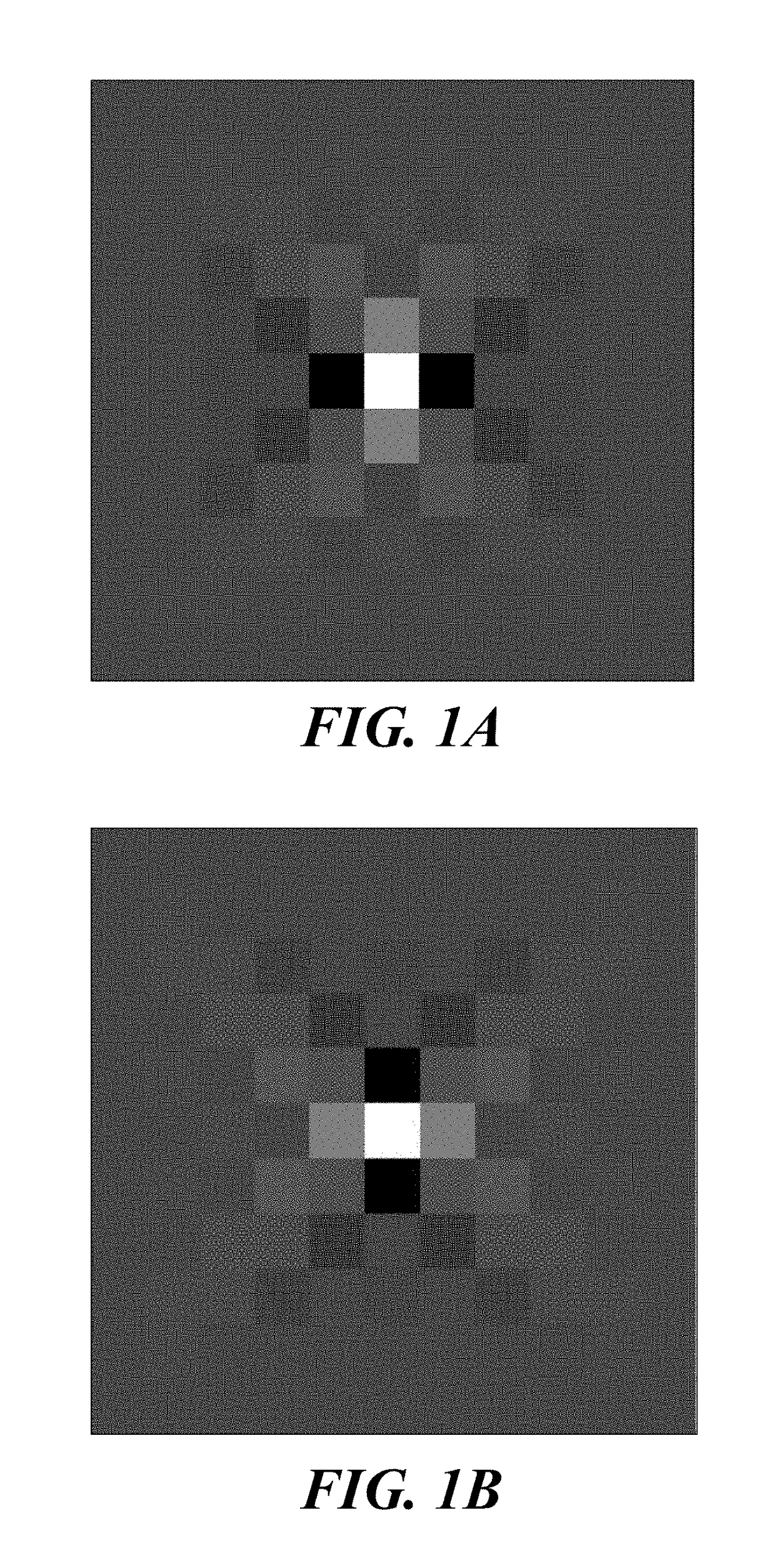
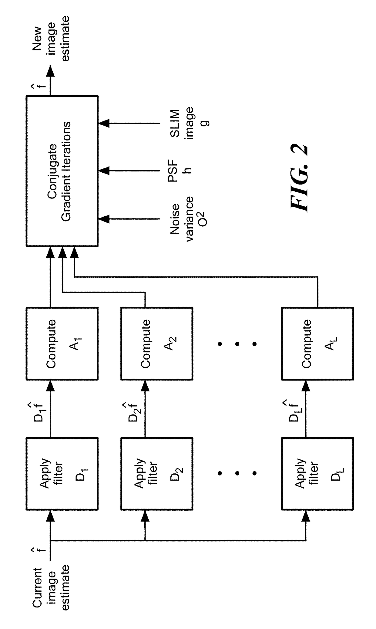
[0081]Application of dSLIM to complex field images obtained by SLIM quantitatively demonstrated the increase in resolution relative to undeconvolved images. All SLIM images were acquired using a white-light source (mean wavelength λ=530 nm); the field of view is 75 μm×100 μm with CCD resolution of 1040×1388. In all reported experiments, the specimen is relatively thin such that the whole image is in focus, and the degradation in the image is only due to a planar PSF. The PSF, depicted in Fig. (a), is obtained experimentally by imaging a sub-resolution 200 nm microbead treated as a point-source. Due to the high SNR provided by SLIM, this PSF closely matches the actual optical transfer function of the imager.

[0082]In all images obtained, the noise level was estimated within the range 10−7-10−6 (for a maximum signal value of 1), which is used as the value of the parameter σ2. The NAs of the objective and condenser are NAo=0.75 and NAc=0.55, respectively.

[0083]The experimentally measure...


PUM


Abstract

Methods and a computer program product for applying deconvolution to spatial light interference microscopy for resolution enhancement with respect to the diffraction limit in two and three dimensions. By exploiting the sparsity properties of the phase images, which is prominent in many biological imaging applications, and modeling of the image formation via complex fields, the very fine structures can be recovered which were blurred by the optics. The resolution improvement leads to higher accuracy in monitoring dynamic activity over time. Experiments with primary brain cells, i.e. neurons and glial cells, reveal new subdiffraction structures and motions. This new information can be used for studying vesicle transport in neurons, which may shed light on dynamic cell functioning. Finally, the method may flexibly incorporate a wide range of image models for different applications and can be utilized for all imaging modalities acquiring complex field images.

Description

[0001]The present application claims priority from U.S. Provisional Application Ser. No. 61 / 653,460, filed May 31, 2012, which is hereby incorporated by reference herein.[0002]This invention was made with Government support under Grant Nos. CBET-0939511, CBET 08-46660 CAREER, CBET-1040462 MRI, 082265 (Physics Frontiers Center: Center for the Physics of Living Cells), and CCF-0916953, all awarded by the National Science Foundation, and under Grant No. R21 CA147967-01, awarded by the National Cancer Institute, and Grant R01GM082837 awarded by the National Institutes of Health. The Government has certain rights in the invention.TECHNICAL FIELD OF THE INVENTION[0003]The present invention pertains to methods for three-dimensional imaging, and, more particularly, for imaging at a resolution greater than the diffraction limit.BACKGROUND OF THE INVENTION[0004]Classical light microscopy techniques cannot be used directly in imaging most biological structures, as such structures are typically...

Claims


Application Information

Patent Timeline
Patent Type & Authority Patents(United States)
IPC IPC(8): G02B21/36G02B21/14G02B21/00
CPCG02B21/361G02B21/008G02B21/14G02B21/365
Inventor BABACAN, S. DERINMIR, MUSTAFA A. H.POPESCU, GABRIELWANG, ZHUO
Owner THE BOARD OF TRUSTEES OF THE UNIV OF ILLINOIS
Why Eureka
Try Eureka