Integration of T2-preparation and motion tracking in magnetic resonance imaging

a magnetic resonance imaging and motion tracking technology, applied in the field of medical imaging, can solve the problems of large imaging delay, small acquisition portion of all necessary imaging data, and often inevitable delay

Active Publication Date: 2017-10-24
THE JOHN HOPKINS UNIV SCHOOL OF MEDICINE
View PDF6 Cites 2 Cited by
  • Summary
  • Abstract
  • Description
  • Claims
  • Application Information

AI Technical Summary

Problems solved by technology

In practice, however, a delay is often inevitable due to other preparations necessary before imaging (e.g. fat saturation, conventional motion tracking using pencil-beam navigator, discussed below).
Usually one can only acquire a small portion of all necessary imaging data (a “segment” of k-space) due to a large variety of physiological and hardware limitations in acquisition speed.
This causes a significant imaging delay during which the fresh T2 contrast degrades for a duration comparable to that of the imaging window itself.
However, this would degrade the quality of motion information obtained with the respiratory navigator, as the interval between the measurement of motion and actual imaging would increase significantly with possible detrimental effects for imaging.
In the standard design of T2 Prep, however, no imaging data or motion data is collected during T2 Prep.
This amounts to a dead time comparable to the duration of the imaging window itself.
Measuring such magnetizations for imaging would not yield data consistent with the rest of the imaging data, leading to artifacts and unacceptable image quality.
MRI is a relatively slow imaging modality and motion of the sample or the subject causes significant image artifacts.
Because respiratory motion in the abdomen is pervasive and nonrigid, complex measurements of the motion field is required if motion is to be corrected.
It is a highly challenging region due to its constant motion, which is generally a mix of two parts: intrinsic cardiac motion and respiratory motion.
This is a significant price to pay for the readout of a single point of respiratory position.
Though multiple navigators can be applied and used for more complex (e.g. affine) motion correction, the delay time due to multiple navigators can be prohibitive for maintaining T2 contrast.
Although during T2 Prep the transverse magnetizations are in flux and unsuitable for imaging, it is noteworthy that the magnetization is still suitable for motion tracking.
Because motion tracking in MRI operates on much less information than image formation, it usually requires lower signal quality and less data.

Method used


Image

  • Integration of T2-preparation and motion tracking in magnetic resonance imaging
  • Integration of T2-preparation and motion tracking in magnetic resonance imaging
  • Integration of T2-preparation and motion tracking in magnetic resonance imaging

Examples


Embodiment Construction

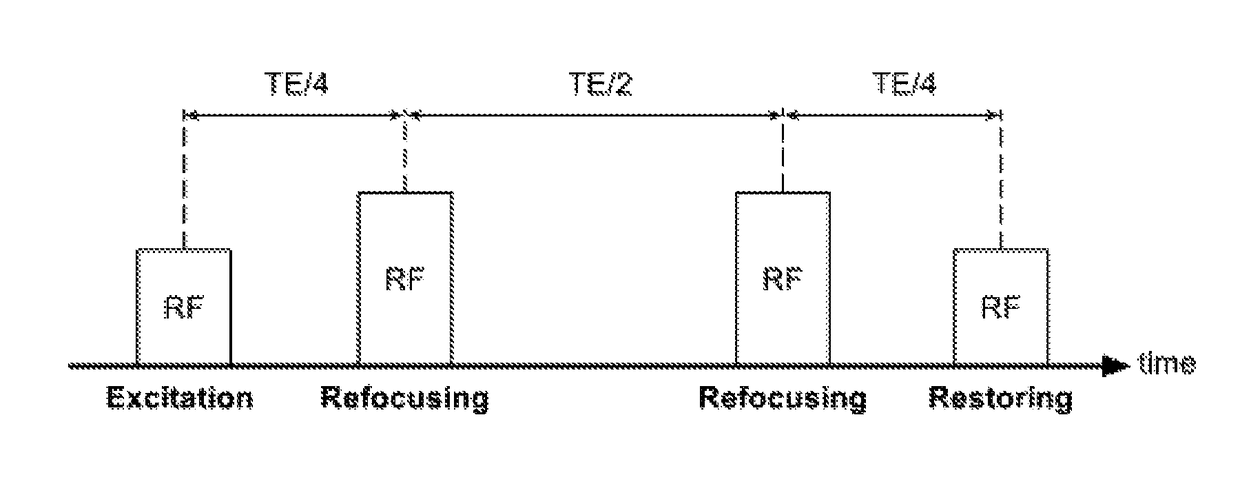
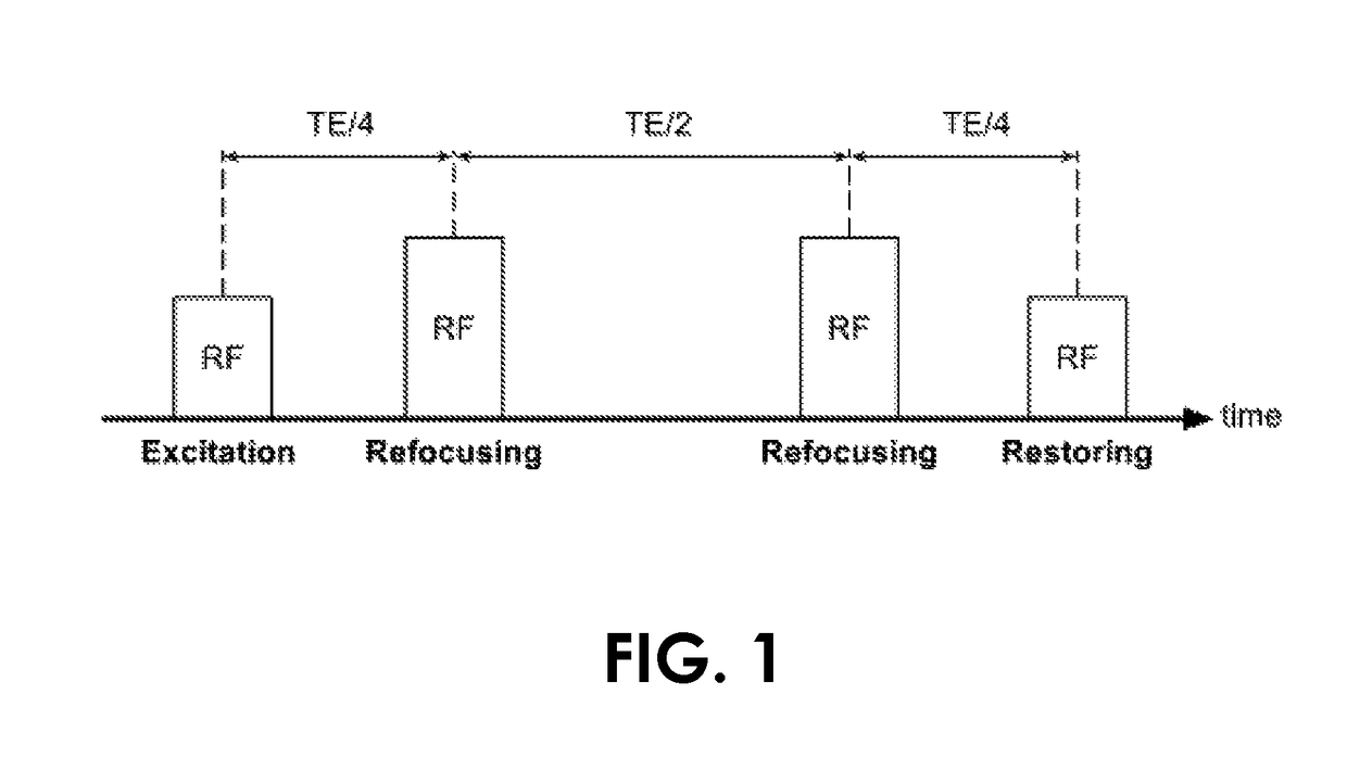
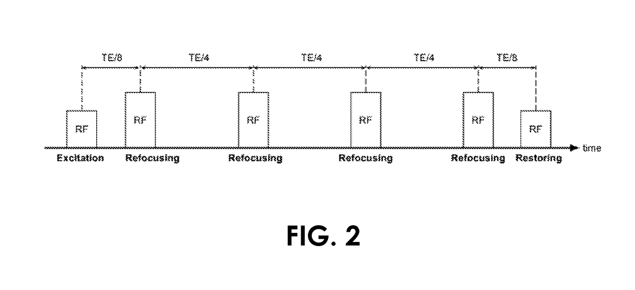
[0030]The presently disclosed subject matter now will be described more fully hereinafter with reference to the accompanying Drawings, in which some, but not all embodiments of the inventions are shown. Like numbers refer to like elements throughout. The presently disclosed subject matter may be embodied in many different forms and should not be construed as limited to the embodiments set forth herein; rather, these embodiments are provided so that this disclosure will satisfy applicable legal requirements. Indeed, many modifications and other embodiments of the presently disclosed subject matter set forth herein will come to mind to one skilled in the art to which the presently disclosed subject matter pertains having the benefit of the teachings presented in the foregoing descriptions and the associated Drawings. Therefore, it is to be understood that the presently disclosed subject matter is not to be limited to the specific embodiments disclosed and that modifications and other ...


PUM


Abstract

An embodiment in accordance with the present invention provides concurrent measurement of motion during T2-weighted magnetic resonance imaging. The present invention combines T2 preparation, a module used to impart T2 contrast, and motion measurement, tracking, and / or correction. The present invention provides for the expedition of more efficient motion compensation during T2-weighted imaging. The proposed invention can be used to provide a variety of measurements of motion, with no overhead in imaging time. The proposed invention also enables T2 contrast imaging to be executed while a subject is breathing freely, without the additional time cost associated with the standard motion tracking methodologies.

Description

CROSS-REFERENCE TO RELATED APPLICATIONS[0001]This application is a 35 U.S.C. §371 U.S. national entry of International Application PCT / US2014 / 034115 having an international filing date of Apr. 15, 2014, which claims the benefit of U.S. Provisional Application No. 61 / 812,321, filed Apr. 16, 2013, the content of each of the aforementioned applications is herein incorporated by reference in their entirety.FIELD OF THE INVENTION[0002]The present invention relates generally to medical imaging. More particularly, the present invention relates to a method of measuring motion during magnetic resonance imaging.BACKGROUND OF THE INVENTION[0003]Magnetic resonance imaging (MRI) is an indispensible modality in medical imaging primarily for its ability to distinguish among a wide array of human tissues as well as among their pathologies. There is an immense body of MRI methodologies at one's disposal to generate diagnostically meaningful contrast to target the medical conditions in question. In g...

Claims


Application Information

Patent Timeline
Patent Type & Authority Patents(United States)
IPC IPC(8): G06K9/00G01R33/56A61B5/00G06T7/207G06T5/00G06T7/00G01R33/567G01R33/565A61B5/055
CPCG06T5/002A61B5/0055A61B5/055A61B5/721A61B5/7207G01R33/5602G01R33/56509G06T7/0012G06T7/207G01R33/5676G06T2207/30092G06T2207/10088G06T2207/30048
Inventor GUO, LIHENGHERZKA, DANIEL A.
Owner THE JOHN HOPKINS UNIV SCHOOL OF MEDICINE
Why Eureka
Try Eureka