
Bionic nano diagnosis and treatment agent for high-field magnetic resonance imaging and preparation method of bionic nano diagnosis and treatment agent

A nanoparticle and mesoporous polydopamine technology, applied in the field of biomedical materials, can solve the problems of low field strength imaging and can not be used for treatment, achieve good biocompatibility, shorten electron relaxation time, improve diagnosis and treatment effect of effect

Pending Publication Date: 2022-03-25
SUN YAT SEN UNIV
View PDF1 Cites 0 Cited by
  • Summary
  • Abstract
  • Description
  • Claims
  • Application Information

AI Technical Summary

Problems solved by technology

The existing known Ho-based nanostructures are mainly limited to oxides (such as Ho 2 o 3 NPs) and fluorides (such as NaHoF 4 NPs), which only have low-field imaging capability and cannot be used for therapy

Method used


Image

  • Bionic nano diagnosis and treatment agent for high-field magnetic resonance imaging and preparation method of bionic nano diagnosis and treatment agent
  • Bionic nano diagnosis and treatment agent for high-field magnetic resonance imaging and preparation method of bionic nano diagnosis and treatment agent
  • Bionic nano diagnosis and treatment agent for high-field magnetic resonance imaging and preparation method of bionic nano diagnosis and treatment agent

Examples


Embodiment 1

[0042] The preparation of embodiment 1 nanometer therapeutic agent Ho-MPDA

[0043] 1. Preparation of Ho-MPDA nanoparticles:

[0044] (1) Dissolve 90 mg of dopamine hydrochloride and 3 mg of holmium nitrate pentahydrate in 3 mL of ultrapure water at 180 rpm and stir for 24 hours in the dark to obtain holmium chelation-dopamine solution;

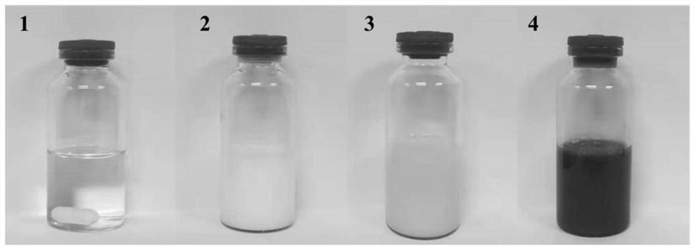
[0045] (2) 36mg surfactant F127 and 0.3mL holmium chelate-dopamine solution are added in the mixed solution of 6mL ethanol and 6mL ultrapure water, 180rpm avoids light and stirs 15min, obtains being colorless transparent solution, such as figure 1 as shown in (1);

[0046] (3) Add 0.1mL of 1,3,5-trimethylbenzene (TMB) solution while shaking in a water bath ultrasonic (4kHz) at 25°C, and continue to ultrasonically disperse for 5 minutes until milky white, such as figure 1 as shown in (2);

[0047] (4) Under magnetic stirring at 180rpm, add 1mL Tris aqueous solution with a concentration of 40mg / mL dropwise, such as figure 1 (3), the solutio...

Embodiment 2

[0051] Preparation of embodiment 2 nanometer therapeutic agent Ho-MPDA

[0052] 1. The procedure is the same as in Example 1, except that the amount of 1,3,5-trimethylbenzene (TMB) is 200 μL.

[0053] 2. Ho-MPDA transmission electron microscope observation:

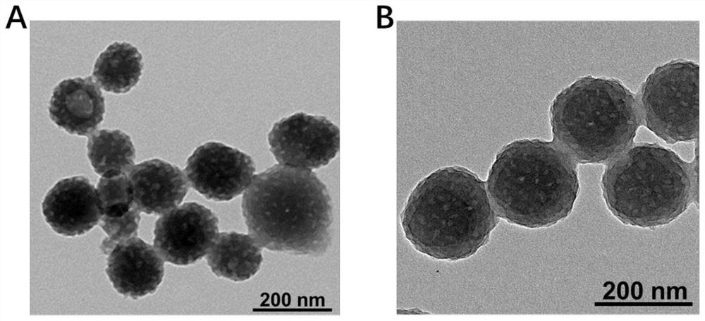
[0054] Prepare the Ho-MPDA prepared in Example 2 into a 100 μg / mL sample solution. After fully ultrasonically dispersed, take 10 μL and drop it on the front of the copper mesh of the carbon support film, place it in a desiccator at room temperature and let it dry naturally. Next, the microscopic morphology of Ho-MPDA was observed using a transmission electron microscope (FEI, Tecnai G2 Spirit). by TEM figure 2 It can be seen from B that the prepared Ho-MPDA is spherical, with a uniform particle size of about 130nm, showing a uniformly distributed mesoporous structure. The shape of the nano-medicine can be controlled by changing the amount of 1,3,5-trimethylbenzene.

Embodiment 3

[0055] The preparation of embodiment 3 nanometer therapeutic agent Ho-MPDA

[0056] The same procedure as in Example 1, except that the amount of 1,3,5-trimethylbenzene (TMB) is 300 μL, and the amount of Tris aqueous solution added is 1.2 mL. The prepared Ho-MPDA is spherical, with a uniform particle size of about 130nm, showing a uniformly distributed mesoporous structure.


PUM

PropertyMeasurementUnit
Specific surface areaaaaaaaaaaa

Abstract

The invention discloses a bionic nano diagnosis and treatment agent for ultrahigh field magnetic resonance imaging and a preparation method thereof, and the nano diagnosis and treatment agent Ho-MPDA is prepared by doping water-soluble dopamine (PDA) with holmium ions (Ho < 3 + >) through a method of doping metal before polymerization. The Ho-MPDA has an obvious T2 contrast enhancement effect under the ultrahigh field intensity of 7.0 T, and the transverse electron relaxation time is obviously shortened. And the transverse relaxation rate T2 (102.71 mM <-1 > S <-1 >) of the Ho-MPDA under the ultrahigh field intensity of 7.0 T is 4.02 times that of a T2 contrast agent (25.52 mM <-1 > S <-1 >), and the imaging effect is greatly improved. Meanwhile, the polydopamine carrier has good biocompatibility, excellent photo-thermal conversion capacity and good light stability, and photo-thermal therapy is achieved under the guidance of ultrahigh-field magnetic resonance imaging.

Description

technical field [0001] The invention relates to the technical field of biomedical materials, in particular to a bionic nano-diagnosis and treatment agent for high-field magnetic resonance imaging and a preparation method thereof. Background technique [0002] Cancer is the main cause of unnatural death cases in all countries in the world, and the global cancer incidence and mortality are increasing year by year. According to the 2020 World Health Organization cancer report, cancer has become the second leading cause of death in the world, and its incidence and mortality have increased by nearly 2040 times. The high mortality rate of cancer is due to the fact that it is mostly diagnosed in the late stage, so early diagnosis and efficient drug treatment are the key to improving the survival rate of cancer patients. [0003] In the diagnosis, treatment and prognosis of cancer, medical imaging technology plays an indispensable role, such as magnetic resonance imaging (MRI), com...

Claims


Application Information

Patent Timeline
IPC IPC(8): A61K49/12A61K49/18A61K41/00A61P35/00B82Y5/00B82Y40/00
CPCA61K49/126A61K49/1857A61K41/0052A61P35/00B82Y5/00B82Y40/00Y02A50/30
Inventor 曹众刘梦
Owner SUN YAT SEN UNIV
Learn More