A kind of preparation method and application of chitosan magnetic nano microsphere
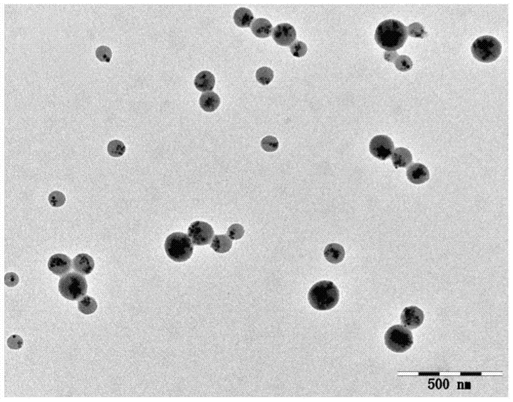
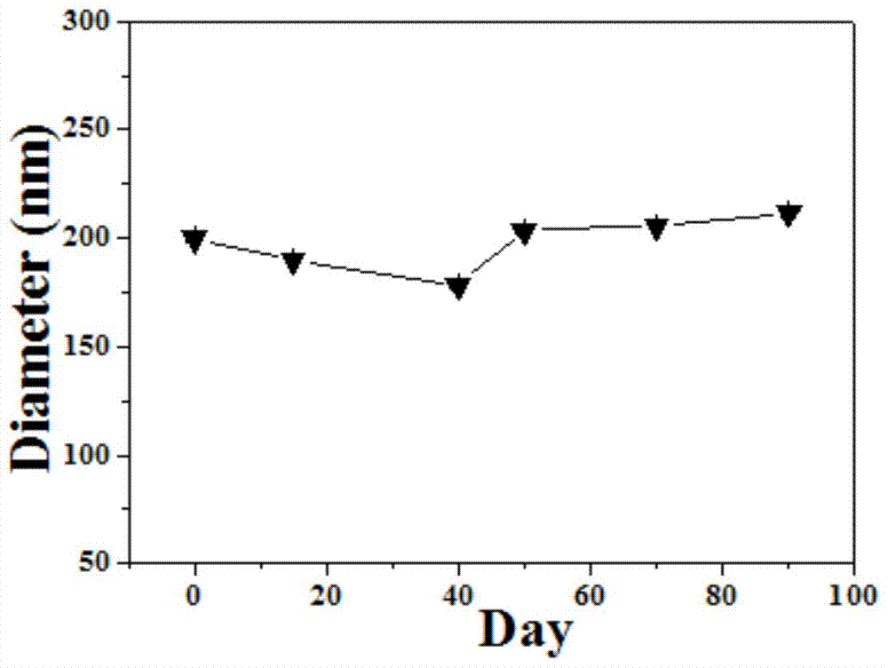
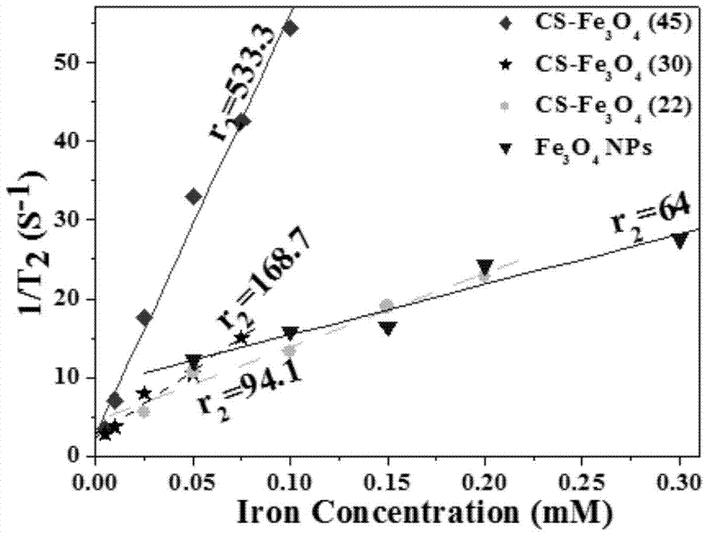
A technology of chitosan microspheres and magnetic nanometers, which is applied in the field of preparation of nanocomposite materials, can solve problems such as difficult control of the particle size distribution of contrast agents, improve diagnostic imaging effects, increase transverse relaxation rate, and reduce in vivo toxicity Effect
- Summary
- Abstract
- Description
- Claims
- Application Information
AI Technical Summary
Problems solved by technology
Method used
Image
Examples
Embodiment 1
[0027] A preparation method of chitosan magnetic nano microspheres, comprising the following steps:
[0028] (1), preparation of superparamagnetic iron oxide-based nanoparticles:
[0029] A mixture of 2.12 g of iron acetylacetonate and 50 ml of tetraethylene glycol was heated at 110° C. for 1 hour under stirring, and then heated and refluxed at 210° C. for 3 hours, under nitrogen protection throughout. The product was separated by ether precipitation at room temperature, washed twice with a mixture of ether and ethanol, and dried to obtain superparamagnetic Fe 3 o 4 Nanoparticles. The product particle size is about 8.8nm.
[0030] (2), chitosan microspheres tightly wrapped superparamagnetic iron oxide nanoparticle clusters:
[0031] Take 0.5 grams of water-soluble chitosan and dissolve it in 80 milliliters of distilled water, add ethylenediamine tetraacetic acid, the mass is 1 / 2 of chitosan, inject 20 milliliters after dissolving, and disperse 0.1 grams of Fe 3 o 4Aqueou...
Embodiment 2
[0034] A preparation method of chitosan magnetic nano microspheres, comprising the following steps:
[0035] (1), preparation of superparamagnetic iron oxide-based nanoparticles:
[0036] A mixture of 2.12 g of iron acetylacetonate and 50 ml of tetraethylene glycol was heated at 110° C. for 1 hour under stirring, and then heated and refluxed at 230° C. for 3 hours, under nitrogen protection throughout. The product was separated by ether precipitation at room temperature, washed twice with a mixture of ether and ethanol, and dried to obtain superparamagnetic Fe 3 o 4 Nanoparticles. The product particle size is about 8.8nm.
[0037] (2), chitosan microspheres tightly wrapped superparamagnetic iron oxide nanoparticle clusters:
[0038] Take 0.5 grams of water-soluble chitosan and dissolve it in 80 milliliters of distilled water, add ethylenediamine tetraacetic acid, the mass is 1 / 3 of chitosan, inject 20 milliliters after dissolving, and disperse 0.1 grams of Fe 3 o 4 Aqueo...
Embodiment 3
[0041] A preparation method of chitosan magnetic nano microspheres, comprising the following steps:
[0042] (1), preparation of superparamagnetic iron oxide-based nanoparticles:
[0043] A mixture of 0.7 g of manganese acetylacetonate, 1.41 g of iron acetylacetonate and 50 ml of tetraethylene glycol was heated at 110° C. for 1 hour under stirring, and then heated and refluxed at 240° C. for 3 hours, under nitrogen protection throughout. The product was separated by ether precipitation at room temperature, washed twice with a mixture of ether and ethanol, and dried to obtain superparamagnetic MnFe 2 o 4 Nanoparticles. The product particle size is about 6.5nm.
[0044] (2), chitosan microspheres tightly wrapped superparamagnetic iron oxide nanoparticle clusters:
[0045] Take 0.5 grams of water-soluble chitosan and dissolve it in 80 milliliters of distilled water, add ethylenediamine tetraacetic acid, the quality is 1 / 5 of chitosan, inject 20 milliliters after dissolving to...
PUM
| Property | Measurement | Unit |
|---|---|---|
| particle diameter | aaaaa | aaaaa |
| particle diameter | aaaaa | aaaaa |
| particle diameter | aaaaa | aaaaa |
Abstract
Description
Claims
Application Information
 - R&D
- Intellectual Property
- Life Sciences
- Materials
- Tech Scout
- Unparalleled Data Quality
- Higher Quality Content
- 60% Fewer Hallucinations
Browse by: Latest US Patents, China's latest patents, Technical Efficacy Thesaurus, Application Domain, Technology Topic, Popular Technical Reports.
© 2025 PatSnap. All rights reserved.Legal|Privacy policy|Modern Slavery Act Transparency Statement|Sitemap|About US| Contact US: help@patsnap.com


