Methods and systems for analysis of single cells
a single cell and analysis method technology, applied in the field of single cell analysis, can solve the problems of lung tissue, hospitalization and death, morbidity and mortality of a particular organism,
- Summary
- Abstract
- Description
- Claims
- Application Information
AI Technical Summary
Benefits of technology
Problems solved by technology
Method used
Image
Examples
example 1
of Gene Expression in Single Cells
[0642]A significant fraction of murine breast CSCs contain relatively low levels of ROS, and so it was hypothesized that these cells may express enhanced levels of ROS defenses compared to their NTC counterparts.
[0643]Single Cell Gene Expression Analysis.
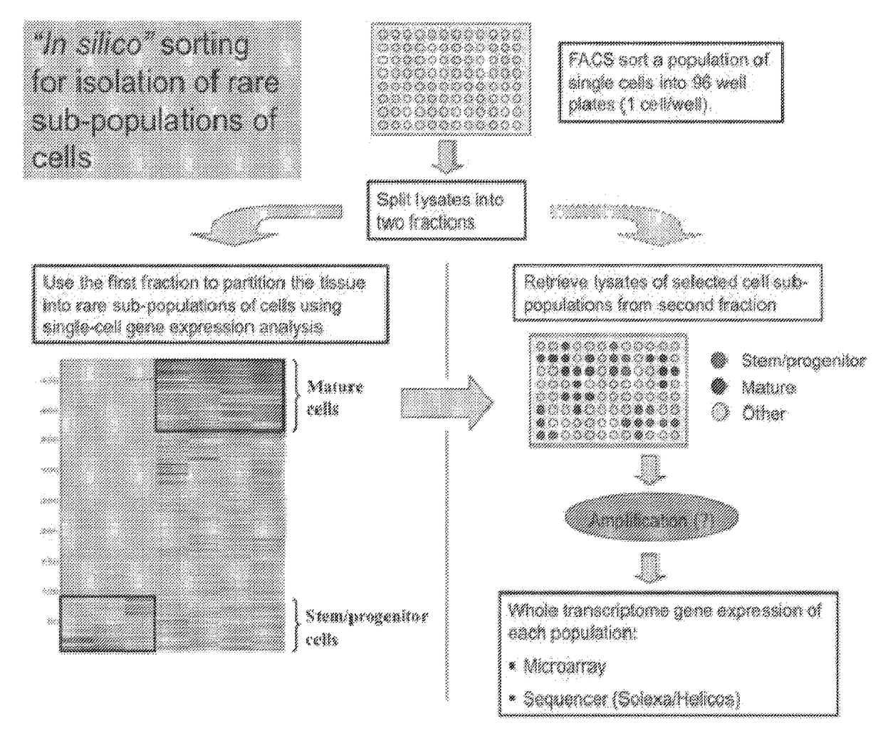
[0644]For the single cell gene expression experiments we used qPCR DynamicArray microfluidic chips (Fluidigm). Single MMTV-Wnt-1 Thy1+CD24+Lin− CSC-enriched cells (TG) and “Not Thy1+CD24+″ Lin” non-tumorigenic (NTG) cells were sorted by FACS into 96 well plates containing PCR mix (CellsDirect, Invitrogen) and RNase Inhibitor (Superaseln, Invitrogen). After hypotonic lysis we added RT-qPCR enzymes (SuperScript III RT / Platinum Taq, Invitrogen), and a mixture containing a pool of low concentration assays (primers / probes) for the genes of interest (Gclm-Mm00514996_m1, Gss-Mm00515065 m 1, Foxo1-Mm00490672_m1, Foxo4-Mm00840140_g1, H ifla Mm00468875_m1, Epas1-Mm00438717 m1). Reverse transcription (15 minut...
example 2
n and Imaging of Human Breast Cancer Xenograft Models with Pulmonary Metastases
[0651]Patient-derived breast cancer specimens (chunks or TICs) were orthotopically transplanted into the mammary fat pads of NOD-SCID mice. Six xenograft tumor models were generated (1 ER+, 1 Her2+ and 4 triple negative ER-PR-Her2−). All four of the triple negative xenografts developed spontaneous lung micro-metastases, demonstrated by IHC stainings, i.e H&E, proliferation marker Ki67 and Vimentin (Vim) stainings. These data suggested that upon implantation into immunodeficent mice, breast tumor cells or TICs are able to adapt to the mouse microenvironment and recapitulate human tumor growth and progression with spontaneous lung metastasis.
[0652]To facilitate dynamic and semi-quantitative imaging of human breast cancer and metastasis in mice, the breast TICs were transduced with firefly luciferase-EGFP fusion gene via the pHRuKFG lentivirus (moi 50) 4 days after implantation, TICs at the primary site were...
example 3
y and Real-Time PCR Analysis of Human Breast MTICs
[0653]Human breast primary tumor initiating cells (TICs) or metastatic TICs (MTICs) (CD44+CD244I′ESA+lineage) were isolated from breast cancer primary site or pleural effusions. Once lung mets were detected in xenograft models, I dissociated lungs with blenzyme (Roche) and stained cells with mouse H2K and human CD44, CD24 and ESA for purifying MTIC populations (CD44′CD2441″ESA+H2K−, FIG. 3a), which grow orthotopic tumors at a ratio of ⅝ with 200-1000 sorted cells, after transplanted into mouse mammary fat pads.
[0654]Shown by microarray analysis and real-time PCR, HIF1a and HIF1 regulated target genes were differentially expressed in MTICs compared to non-tumorigenic tumor cells, including Snail, Zeb2, Vimentin, E-cadherin, Lox, Cox2, VEGF, etc. (FIG. 3B). Co-localization of HIFI a, Vimentin and CD44 were confirmed by immunohistochemistry staining.
PUM
| Property | Measurement | Unit |
|---|---|---|
| volume | aaaaa | aaaaa |
| volume | aaaaa | aaaaa |
| volume | aaaaa | aaaaa |
Abstract
Description
Claims
Application Information
 - R&D
- Intellectual Property
- Life Sciences
- Materials
- Tech Scout
- Unparalleled Data Quality
- Higher Quality Content
- 60% Fewer Hallucinations
Browse by: Latest US Patents, China's latest patents, Technical Efficacy Thesaurus, Application Domain, Technology Topic, Popular Technical Reports.
© 2025 PatSnap. All rights reserved.Legal|Privacy policy|Modern Slavery Act Transparency Statement|Sitemap|About US| Contact US: help@patsnap.com


