Systems and methods for rapid neural network-based image segmentation and radiopharmaceutical uptake determination

a neural network and image segmentation technology, applied in the field of systems and methods for rapid neural network-based image segmentation and radiopharmaceutical uptake determination, can solve the problems of patient's family likely having questions, patient may very well struggle to make sense of this information, and the process is limited, so as to improve computational efficiency, computational efficiency, and computational efficiency.

Active Publication Date: 2019-07-11
EXINI DIAGNOSTICS +1
View PDF0 Cites 41 Cited by
  • Summary
  • Abstract
  • Description
  • Claims
  • Application Information

AI Technical Summary

Benefits of technology

[0019]The ‘bounding box’ approach of identifying one or more portions of a 3D anatomical image that is/are relevant to the analysis at hand, e.g., prior to applying a second CNN (for detailed segmentation), improves computational efficiency by removing a large portion of the initial 3D anatomical image prior to the more computationally intensive subsequent processing. This approach is more computationally efficient than performing the detailed segmentation on the entire initial 3D anatomical image since, for example, identification of the pelvic region (e.g., the vertices of a cuboid bounding box) is simpler than detailed segmentation of a prostate, bladder, and/or other tissues of interest. Not only is this approach more computationally efficient, it also may result in more accurate subsequent processing, e.g., the more detailed segmentation provided by the second CNN. This is because, for example, 3D anatomical images obtained at different medical institutions using different machines vary in size (e.g., where varying size means there are different numbers of voxels of the images, and/or different volumes of the patient tissue represented in the images), and training the second CNN for automated detailed segmentation of the prostate using portions of 3D anatomical training images having a more standardized image volume size, and within which the organs and other tissue regions of interest lie, results in a more robust, accurate segmentation.
[0020]Another advance described herein that improves the performance of the automated measurement of uptake metrics in the prostate (or, more broadly, a particular organ or tissue region of interest) via this multi-image approach (i.e., analysis of a 3D anatomical image in combination with a 3D functional image) is the accurate identification of one or more tissue regions in addition to the prostate, and the accounting for imaging agent uptake in those regions in the determination of (i) uptake metrics in the prostate and/or (ii) an identification and/or staging of prostate cancer. Certain imaging agents comprising a PSMA binding agent have high uptake in certain organs, which may affect the identification of diseased tissue (e.g., prostate cancer). For example, uptake of a radionuclide labelled PSMA binding agent by the bladder may result in scattering in the 3D functional image, and may reduce accuracy of the measured imaging agent intensity in the prostate, which is located ne

Problems solved by technology

The process may also involve repeating the imaging study due to inconclusive results, or ordering further tests based on initial results.
There are limitations associated with this process, both from the perspective of the physician and from the perspective of the patient.
The physician may show the patient his PET/SPECT images and may tell the patient a numerical risk associated with various treatment options or likelihood of

Method used


Image

  • Systems and methods for rapid neural network-based image segmentation and radiopharmaceutical uptake determination
  • Systems and methods for rapid neural network-based image segmentation and radiopharmaceutical uptake determination
  • Systems and methods for rapid neural network-based image segmentation and radiopharmaceutical uptake determination

Examples


example 4

J. Selection of TBR Threshold for Clinically Significant Findings

[0327]Example 4 is an example showing how a TBR threshold value for partitioning patient prostate cancer pathology into clinically significant and clinically non-significant classifications can be determined.

[0328]Two datasets of SPECT / CT images were combined to select an appropriate threshold value. A first dataset comprised images of healthy individuals, taken from a phase I study of the 1404 drug. This dataset contained originally 14 images. Segmentation of a prostate within the images was performed in accordance with the approaches described herein and two images where segmentation of the prostate clearly failed were excluded, resulting in 12 remaining images. A second data set comprised images of individuals with prostate cancer, originating from a phase II study of the 1404 drug. The images were partitioned based on the subject's Gleason grades on histopathology from radical prostatectomy. A total Gleason Score ...

example 5

K. Application of AI to Phase 3 Study MIP-1404-3301 (“proSPECT-AS”)

[0334]MIP1404-3301 is a pivotal phase 3 multicenter trial of 99mTc-MIP1404 Injection for the detection of prostate cancer. MIP-1404-3301 is titled “A Phase 3 Study to Evaluate the Safety and Efficacy of 1404 SPECT / CT Imaging to Detect Clinically Significant Prostate Cancer in Men with Biopsy Proven Low-Grade Prostate Cancer who are Candidates for Active Surveillance”. Cohort A patients were men with biopsy-proven low to intermediate grade prostate cancer (Gleason score 3+3 or 3+4) who were candidates for active surveillance but elected to have radical prostatectomy. Cohort B patients were biopsy-proven very low risk prostate cancer patients who scheduled to undergo routine re-biopsy as part or routine active surveillance. The phase 3 study was initiated in December 2015, and enrollment is now complete. The computer-assisted diagnosis (CADx) software device is to be used as the primary reading methodology to analyze ...

example 6

L. Training and Validation of Convolutional Neural Networks Implemented by the First Machine Learning Module (Localization Machine) and Second Machine Learning Module (Segmentation Machine)

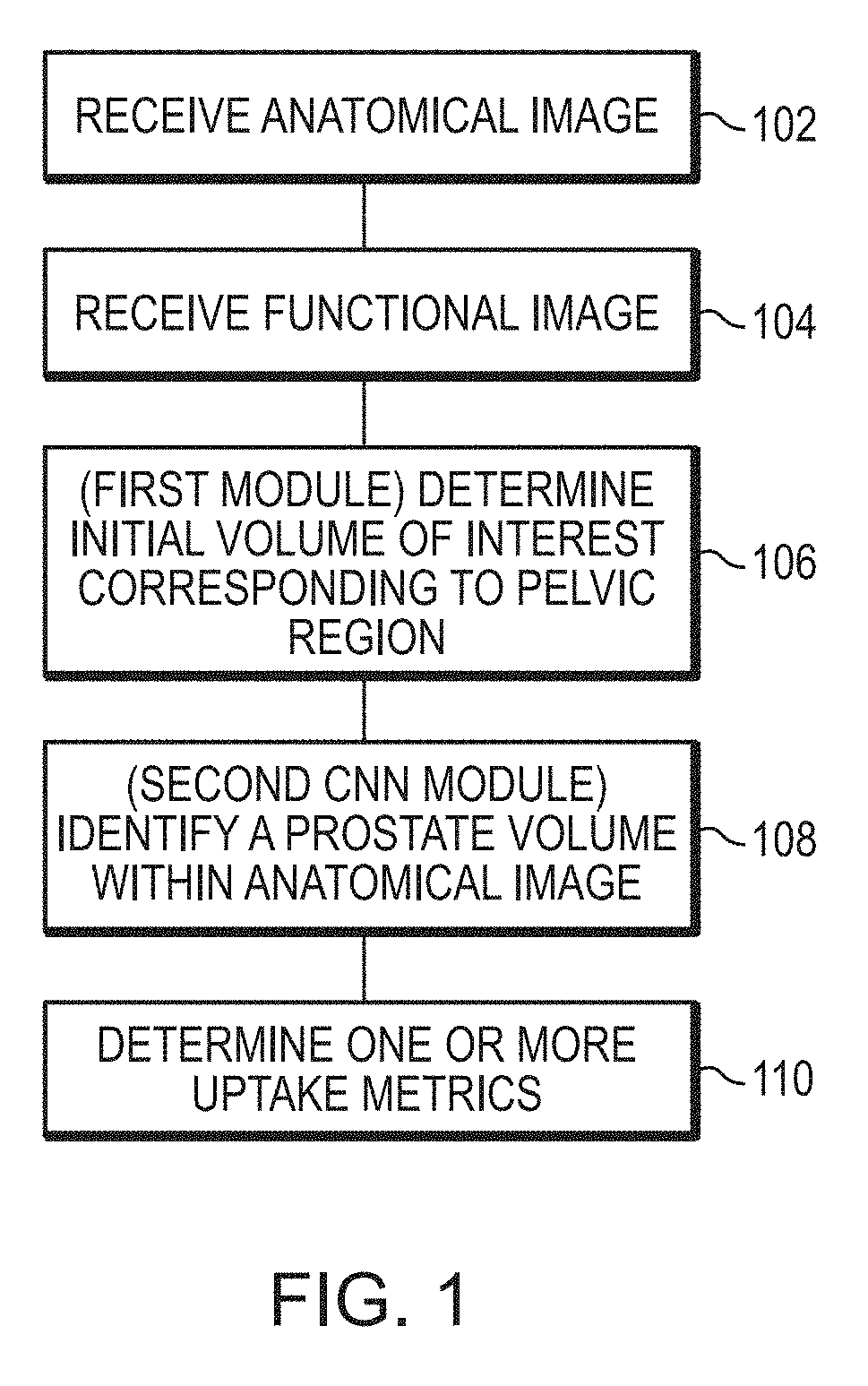
[0339]Example 5 is an example showing training and validation of CNN modules used to segment CT images to identify various tissue volumes, including a prostate volume, in accordance with the aspects and embodiments described herein. In this example, the neural networks were defined and trained using the Keras framework with the Tensorflow backend.

[0340]i. Training and Validation Data

[0341]The training and validation data comprised CT images coupled with semi-automated segmentations corrected by a radiologist delineating all or some of the following body parts: (i) a prostate; (ii) a urinary bladder; (iii) a rectum; (iv) a left gluteus maximus; (v) a right gluteus maximus; (vi) a left hip bone; (vii) a right hip bone; and (viii) a sacrum and coccyx

[0342]To train a localization CNN, 90 high quality...


PUM


Abstract

Presented herein are systems and methods that provide for automated analysis of three-dimensional (3D) medical images of a subject in order to automatically identify specific 3D volumes within the 3D images that correspond to specific organs and/or tissue. In certain embodiments, the accurate identification of one or more such volumes can be used to determine quantitative metrics that measure uptake of radiopharmaceuticals in particular organs and/or tissue regions. These uptake metrics can be used to assess disease state in a subject, determine a prognosis for a subject, and/or determine efficacy of a treatment modality.

Description

CROSS REFERENCE TO RELATED APPLICATIONS[0001]This application claims priority to and benefit of U.S. Provisional Application No. 62 / 614,935, filed Jan. 8, 2018, the content of which is hereby incorporated by reference in its entirety.FIELD OF THE INVENTION[0002]This invention relates generally to methods, systems, and architectures for automated analysis and / or presentation of medical image data. More particularly, in certain embodiments, the invention relates to automated identification of one or more particular regions of interest (e.g., corresponding to specific organs or tissue) within images of a subject and determination of radiopharmaceutical uptake within such region(s), e.g., for identification and / or staging of disease, e.g., prostate cancer.BACKGROUND OF THE INVENTION[0003]Targeted image analysis involves the use of radiolabeled small molecules that bind to specific receptors, enzymes and proteins in the body that are altered during the evolution of disease. After adminis...

Claims


Application Information

Patent Timeline
IPC IPC(8): A61B6/00G06T15/08G06T7/00G06T5/30G06K9/46A61B6/03A61K51/04G06V10/764
CPCA61B6/5217G06T15/08G06T7/0014G06T5/30G06K9/4647A61B6/037A61B6/50A61B6/466A61B6/467A61B6/465A61B6/5223A61B6/5258A61B6/5229A61K51/0478G06T2207/30081G06T2207/30096G06T2207/10108G06K2209/051G06T2200/24G06F3/04842G06N3/04G06N3/0454G06N3/08G06T7/143G06T7/11G06T7/0012G06T2207/10104G06T2207/20081G06T2207/10081G06T2207/10088G06T2207/30008G06N3/082G16H50/30G06T2207/20084G06V20/64G06V2201/031G06V10/82G06V10/764G06N3/045
Inventor SJOSTRAND, KARL VILHELMRICHTER, JENS FILIP ANDREASJOHNSSON, KERSTIN ELSA MARIAGJERTSSON, ERIK KONRAD
Owner EXINI DIAGNOSTICS
Why Eureka
Try Eureka