Compounds, kits and methods for use in medical imaging

A technology of kits and imaging agents, applied in the field of new compounds for medical imaging and treatment, which can solve problems such as blurring

Inactive Publication Date: 2007-09-12
KONINKLIJKE PHILIPS ELECTRONICS NV
View PDF0 Cites 4 Cited by
  • Summary
  • Abstract
  • Description
  • Claims
  • Application Information

AI Technical Summary

Problems solved by technology

Additionally, accumulation in clearance pathways such as the hepatic bile ducts or kidneys m

Method used


Image

  • Compounds, kits and methods for use in medical imaging
  • Compounds, kits and methods for use in medical imaging
  • Compounds, kits and methods for use in medical imaging

Examples


Embodiment 1

[0066] Example 1: Pretargeted Imaging of Tumors Using Azide-Glucose

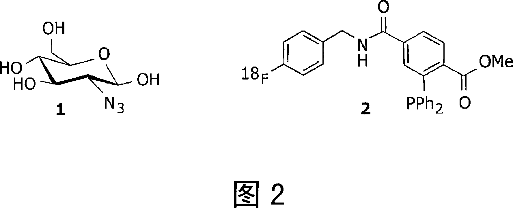
[0067] Refer to Figure 2.

[0068] Systemic administration of azide-glucose probe 1. Following optimal accumulation in tissues with high glucose uptake (such as tumors) and optimal clearance from non-target tissues and blood, administration of 18 F labeled imaging probe 2. Construct 2 is intracellularly conjugated to entrapped 1 via a Staudinger ligation. After clearing of unbound 2, PET images can be recorded, delineating tumor location and activity.

Embodiment 2

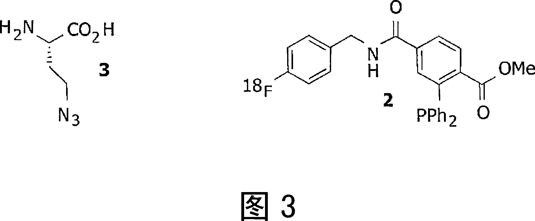
[0069] Example 2: Pretargeted Imaging of Tumors Using Azide-Amino Acids

[0070] Refer to Figure 3. The E. coli translation apparatus [bertozzi_PNAS2002] recognizes Azidohomoalanine (3) as a methionine substitute, which can serve as a metabolic / proliferation marker. Systemic administration of azidohomoalanine (3). Following optimal accumulation in tissues with high amino acid uptake (such as tumors) and optimal clearance from non-target tissues and blood, administration of 18 F labeled imaging probe 2. Construct 2 is intracellularly conjugated to entrapped 3 via a Staudinger ligation. After clearing of unbound 2, PET images can be recorded, delineating tumor location and activity.

Embodiment 3

[0071] Example 3: Pretargeted Imaging of Tumors Using Azide-Nucleosides

[0072] Refer to Figure 4.

[0073] Cell proliferation is increased in tumors, which leads to increased DNA replication and thus to an increased requirement for nucleosides. Given azide-modified thymidine 4, it is taken up by rapidly dividing cells. After optimal uptake in target cells, inject 18 F-labeled cyclooctyne compound 5, which binds to entrapped 4 via [3+2]azide-alkyne cycloaddition. After clearing of unbound 5, PET images can be recorded, delineating tumor location and activity.


PUM


Abstract

The use of a selective chemical and bioorthogonal reaction providing a covalent ligation such as the Staudinger ligation, in targeted molecular imaging and therapy is presented, more specifically with interesting applications for pre-targeted imaging or therapy. Current pre-targeted imaging is hampered by the fact that it relies solely on natural/biological targeting constructs (i.e. biotin/streptavidin). Size considerations and limitations associated with their endogenous nature severely limit the number of applications. The present invention describes how the use of an abiotic, bio-orthogonal reaction which forms a stable adduct under physiological conditions, by way of a small or undetectable bond, can overcome these limitations.

Description

field of invention [0001] The present invention relates to novel compounds, kits and methods for medical imaging and therapy. In particular the invention relates to FDG, FLT and 11 Alternatives to C Methionine. Background of the invention [0002] The imaging modalities positron emission tomography (PET) and single photon emission computed tomography (SPECT) rely on radiolabeled drugs to produce images. [0003] PET records high-energy gamma-rays emitted from the subject. Frequently used positron-emitting isotopes include 15 O. 13 N. 11 C and 18 F, the latter being used as a substitute for hydrogen. Labeled molecular probes can be introduced into the subject, and PET imaging can follow the distribution and concentration of the injected molecules. SPECT imaging uses radiopharmaceuticals with isotopes that decay under emission of gamma radiation. SPECT enables the visualization of biological processes with kinetics on the scale of hours to days. The most commonly use...

Claims


Application Information

Patent Timeline
IPC IPC(8): A61K49/00A61K51/02A61K47/48
CPCA61K47/48353B82Y5/00A61K51/0491A61K47/665A61P7/02A61P9/00A61P9/10A61P29/00A61P31/04A61P35/00
Inventor 马克·S.·罗比亚尔霍尔格·格吕尔
Owner KONINKLIJKE PHILIPS ELECTRONICS NV
Why Eureka
Try Eureka