Automated immunoanalyzer system for performing diagnostic assays for allergies and autoimmune diseases

an immunoanalyzer and allergy technology, applied in the field of automatic immunoanalyzer system and process for performing diagnostic assays for allergies and autoimmune diseases, can solve the problem that the original particle fraction will be lost in subsequent chemistry processes, and achieve the effect of maximum detection sensitivity

Active Publication Date: 2014-09-18
HYCOR BIOMEDICAL LLC
View PDF10 Cites 25 Cited by
  • Summary
  • Abstract
  • Description
  • Claims
  • Application Information

AI Technical Summary

Benefits of technology

[0017]According to certain specific aspects of the present disclosure, horseradish peroxidase (HRP) conjugated to an anti-IgE antibody can be used as an indirect label when washing the immune complex to remove unbound IgE, particularly as the reaction of PS-Atto with an HRP labeled conjugate generates sustained high-intensity luminescence for maximum detection sensitivity in solution assays.

Problems solved by technology
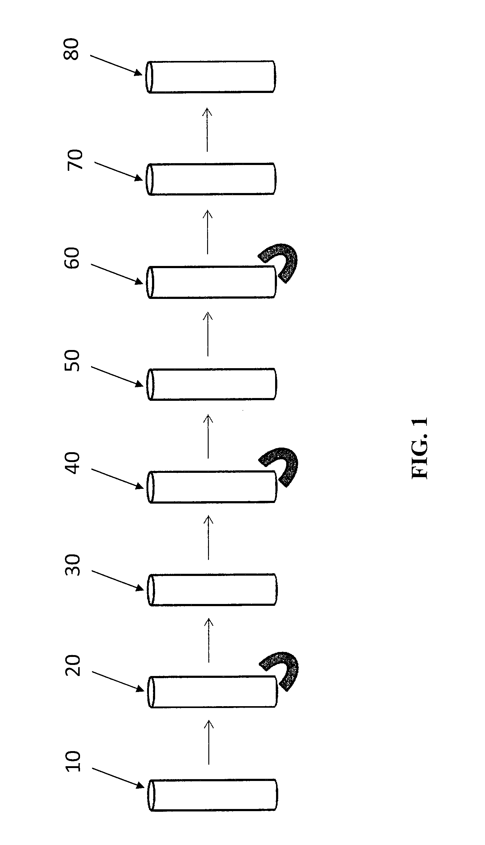
A consequence of these washing steps, however, is that some fraction of the original particles will be lost for subsequent chemistry processes.

Method used


Image

  • Automated immunoanalyzer system for performing diagnostic assays for allergies and autoimmune diseases
  • Automated immunoanalyzer system for performing diagnostic assays for allergies and autoimmune diseases
  • Automated immunoanalyzer system for performing diagnostic assays for allergies and autoimmune diseases

Examples


example 1

Biotinylation of Anti-Human IgE or Allergen Extracts

[0057]2 μL of NHS-PEG12-Biotin (Pierce) 250 mM in DMSO is added to 1 mL of affinity purified anti-human IgE (ImmunoReagents) 5.0 mg / mL in Phosphate Buffered Saline (PBS). Or, 1.6 μL of NHS-PEG12-Biotin (Pierce) 250 mM in DMSO is added to 1 mL of allergen extracts 1.0 mg / mL in Phosphate Buffered Saline (PBS).

[0058]The reagent solution is mixed and placed on ice for 2 hours. Free biotin reagent is separated from the biotinylated antibody by dialysis against two changes of PBS (volume ratio of antibody to buffer—1:100) at 2-8° C. for 4 hours and overnight.

example 2

Preparation of Fluo-Bead

[0059]5 μL of Biotin-Fluo (Alexa Fluor 594 Biocytin, Sodium Salt, Life Technologies) 1 mM in ddH2O is added into 45 mL of PBSTHP Buffer (10 mM sodium phosphate, pH 7.4, 0.9% (w / v) NaCl, 0.05% (v / v) Tween-20, 10 mg / mL HSA, 1% (v / v) ProClin 950). Mix well.

[0060]5 mL of SA-Speed Bead (Sera-mag Speedbeads Streptavidin-Coated Magnetic Particles, Thermo) 10 mg / mL is added into the Biotin-Fluo solution and mixed well.

example 3

Assay for Specific IgE Levels to Allergens

[0061]10 μL Fluo-Bead (Fluorescence labeled para-magnetic microparticles) at bead concentration 1 mg / mL is dispensed into the reaction cuvette; 40 μL of biotin-allergen (e.g., Egg white, Milk, Peanut, etc) or biotin-anti-IgE antibody, is dispensed and mixed into the Fluo-Bead, and incubated for 1-10 min at 37° C. After washing, allergen- or anti-IgE-coated beads are resuspended in 40 μL of reaction buffer. Serum samples obtained from atopic and non-atopic individuals are assayed against allergens. A 10 μL sample was added to 40 μL of suspended allergen-coated beads in reaction cuvette. For the six point standard curve, 10 μL of serum standards (secondary standards calibrated against the WHO IgE Standard 75 / 502) are each added to 40 μL of anti-IgE-coated beads in a reaction cuvette. While various different labeled anti-IgE conjugates can be utilized in accordance with the present teachings, in accordance with certain teachings, the following ...


PUM

PropertyMeasurementUnit
pHaaaaaaaaaa
pHaaaaaaaaaa
pHaaaaaaaaaa

Abstract

A quantitative method for performing an automated diagnostic assay, comprising: incubating a capture reagent with a streptavidin-coated medium to form a solid phase complex; washing the solid phase complex to remove excess capture reagent; incubating the solid phase complex with a serum sample to form an immune complex; washing the immune complex to remove any unbound sample; incubating the immune complex with a conjugate to create an immune-conjugate complex; washing the immune-conjugate complex to remove any unbound conjugate; introducing a substrate capable of generating a quantifiable response; and calibrating the response generated from introducing the substrate.

Description

CROSS-REFERENCE TO RELATED APPLICATIONS[0001]This application is related to and claims priority to U.S. Provisional Patent Application Ser. Nos. 61 / 791,295 and 61 / 791,879, each of which were filed on Mar. 15, 2013, the complete and entire disclosures of which are hereby expressly incorporated by reference herein.TECHNICAL FIELD[0002]The present teachings are related to a system and process for performing diagnostic assays, and more particularly to an automated immunoanalyzer system and process for performing diagnostic assays for allergies and autoimmune diseases.BACKGROUND OF THE DISCLOSURE[0003]The statements in this section merely provide background information related to the present disclosure and should not be construed as constituting prior art.[0004]During an automated immunochemistry analysis, analyte molecules in a patient's biological sample (e.g., serum or plasma) attach to paramagnetic particles. To remove background signals associated with potential chemical sources tha...

Claims


Application Information

Patent Timeline
Patent Type & Authority Applications(United States)
IPC IPC(8): G01N33/564G01N33/543G01N33/68
CPCG01N35/1011G01N2035/0453G01N2021/6484G01N2035/1062G01N33/54393G01N33/564G01N33/6854G01N33/5306G01N33/54326G01N33/582G01N33/6893G01N35/00G01N2800/24G01N35/0098G01N35/04G01N21/645G01N21/6428G01N21/76G01N33/569G01N2201/062G01N2201/08G01N2333/4703G01N2333/62G01N2333/78G01N21/274G01N2201/12707G01N2201/084G01N2201/0646G01N2201/0469Y10T436/119163G01N33/5434G01N33/5695G01N33/56983
Inventor VAN CLEVE, MARK DAVIDREID, TAYLOR ADDISONTRONDLE, LINDA MARIEHUNG, VICTORIALUO, YIRIEGER, DENNIS EDWINMCMENAMY, EVAN PHILLIPRAGHAVAN, NANDITHARELIFORD, MORKOAH BLAYCANFIELD, DOUGLAS JOHNNIE, YILINGSINSON, EDSEL LAWRENCE NOCHEVANDE WETERING, SCOTT WILLIAMTAYLOR, TERIKNOX, TRAVISCUARESMA JACALNE, FRAN ZYLOWESTON, JAMESBAO-GUEY CHAN, JENNIFERTUVIVO, STEPHANIESCHELL, RACHEL SARAHDIAMOND, RONALD NORMANGANN, STEVE MICHAELHALL, ERIC DARNELLHWANG, TAE HOMORTON, JOHN LEWISMOSKALEV, ANATOLYSTACK, MARINELA GOMBOSEVSARGEANT, BRUCE ALAN
Owner HYCOR BIOMEDICAL LLC
Why Eureka
Try Eureka