Medical imaging method and medical imaging system

A medical imaging system and medical imaging technology, applied in the field of medical imaging methods and medical imaging systems, can solve the problems affecting the accuracy of medical imaging and the lack of system sensitivity, so as to avoid the loss of attenuation information, avoid excessive coverage, and improve accuracy Effect

Active Publication Date: 2017-09-29
SHANGHAI UNITED IMAGING HEALTHCARE
View PDF3 Cites 15 Cited by
  • Summary
  • Abstract
  • Description
  • Claims
  • Application Information

AI Technical Summary

Problems solved by technology

[0004] However, when performing PET image reconstruction, it often occurs that some coincident events cannot be correctly used in PET image reconstruction and have to be discarded, and there are unnecessary parts of the collected coincident events, which will Directly lead to loss of system sensitivity, thus affecting the accuracy of medical imaging

Method used


Image

  • Medical imaging method and medical imaging system
  • Medical imaging method and medical imaging system
  • Medical imaging method and medical imaging system

Examples


Embodiment 1

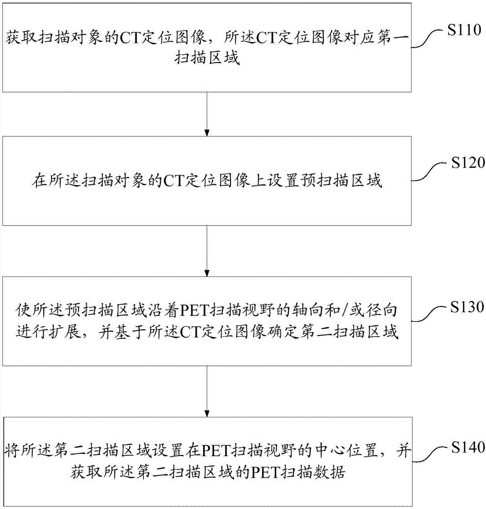
[0053] figure 1 It is a schematic flow chart of the medical imaging method in Embodiment 1 of the present invention, figure 2 It is a schematic diagram of the steps of the medical imaging method in Embodiment 1 of the present invention, Figure 3a with Figure 3b It is a schematic structural diagram during the process of determining the second scanning area in the medical imaging method in Embodiment 1 of the present invention. Combine below figure 1 , figure 2 with Figure 3a - Figure 3b As shown, the medical imaging method in this embodiment will be described in detail.

[0054] First, in step S110, a CT positioning image of the scanning object is acquired, and the CT positioning image corresponds to the first scanning area D1, that is, the first scanning area D1 is the scanning area of ​​the CT positioning image. When subsequently determining the effective area for PET image reconstruction, the CT positioning image is used to determine the boundary of the effectiv...

Embodiment 2

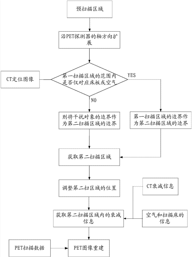
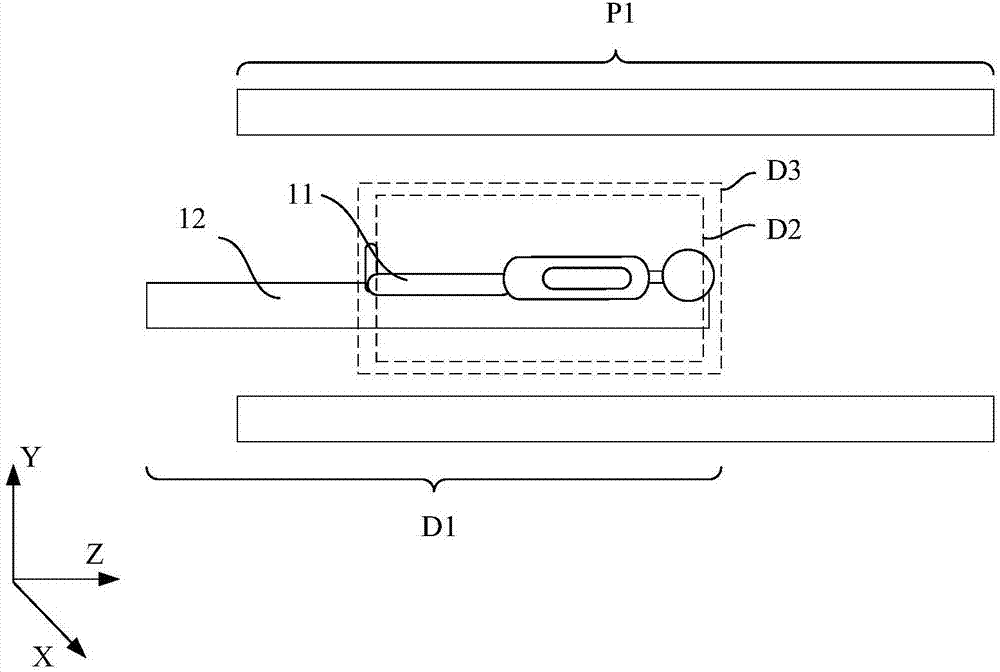
[0095] The difference from Embodiment 1 is that in this embodiment, interference objects other than the bed board and air are corresponding within the area range of the first scanning area D1. specific reference Figure 4a with Figure 4b Shown is a schematic structural diagram during the process of determining the second scanning region in the medical imaging method in Embodiment 2 of the present invention.

[0096] refer to Figure 4a As shown, the area range of the first scanning area includes the scanning object 11 , the bed board 12 , the air and the interfering object 14 , therefore, the first scanning area cannot be directly defined as the second scanning area. After the CT scan area D3 is defined, it is necessary to continue to expand the CT scan area D3, and obtain a second scan area based on the CT positioning image.

[0097] Specifically, continue to expand the area boundary toward the axis of the PET scanning field of view, for example:

[0098] First, expand t...

Embodiment 3

[0103] The invention also provides a medical imaging system. Figure 5 It is a schematic structural diagram of the medical imaging system in Embodiment 3 of the present invention, as Figure 5 As shown, the medical imaging system includes:

[0104] Bed board 110, used to carry the scanning object;

[0105] A CT scanning device 120, configured to acquire a CT positioning image of the scanned object;

[0106] A scan area defining unit 130, configured to set a pre-scan area on the CT positioning image, and expand the pre-scan area along the axial and / or radial direction of the PET scan field of view to determine a second scan area;

[0107] The PET detector 150 is configured to set the second scanning area at the center of the PET scanning field of view, and acquire PET scanning data of the second scanning area.

[0108] In addition, the medical imaging system further includes:

[0109] The attenuation information acquiring unit 140 is configured to acquire attenuation inform...


PUM


Abstract

The invention provides a medical imaging method and medical imaging system. The method comprises the steps that a pre-scanning area extends in the axial direction and/or radial direction of the PET scanning visual field, and a second scanning area is determined based on a CT positioning image; the second scanning area is arranged on the center of the PET scanning visual field, and PET scanning data of the second scanning area is obtained. According to the medical imaging method and medical imaging system, combined with the CT positioning image, the range of the second scanning area can be defined to the maximum, and accordingly the collected PET scanning data can be sufficiently used. After the range of the second scanning area is defined, the relative location in the PET axial scanning visual field can be further adjusted, the situation that the relative location of scanning objects is located in the area with higher detection sensitivity, and data collection with maximum sensitivity is achieved.

Description

technical field [0001] The invention relates to the field of medical imaging, in particular to a medical imaging method and a medical imaging system. Background technique [0002] Positron Emission Tomography (PET) is a non-invasive nuclear medicine imaging diagnosis technology widely used clinically. It provides functional information such as metabolism of the living body by imaging the radioactive tracer injected into the living body. It plays an important role in diagnosis, efficacy evaluation, basic medical research and new drug development. [0003] PET is a technique that uses positron nuclide as a tracer to understand the functional metabolic state of the lesion through the uptake of the tracer at the lesion site, and then diagnose the disease. Specifically: the radioactive tracer drug labeled with a positron nuclide is injected into the subject, and the positron nuclide decays and emits positrons, which undergo an annihilation reaction with the negative electrons in...

Claims


Application Information

Patent Timeline
Patent Type & Authority Applications(China)
IPC IPC(8): A61B6/03A61B6/00
CPCA61B6/037A61B6/488A61B6/5211A61B6/032A61B6/4417A61B6/5235A61B6/545G06T2200/04G06T7/33G06T2207/10081G06T2207/10104A61B6/5205G06T11/006G16H50/20G06T7/11G06T7/0012G06T11/003G06T7/13
Inventor 孙友军顾笑悦刘伟平
Owner SHANGHAI UNITED IMAGING HEALTHCARE
Why Eureka
Try Eureka