

Tumor displace mimetism organization chip
A tumor metastasis and tissue chip technology, which is applied in the preparation of material inspection products, test samples, biological tests, etc., can solve the problems of staying, no comparative research on the dynamic changes of cell-related gene expression, etc.
- Summary
- Abstract
- Description
- Claims
- Application Information
AI Technical Summary
Problems solved by technology
Method used
Image

Examples
Embodiment 1
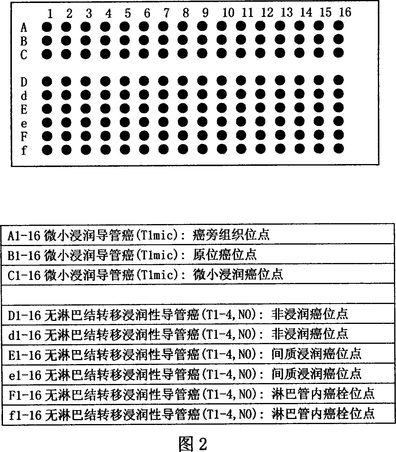
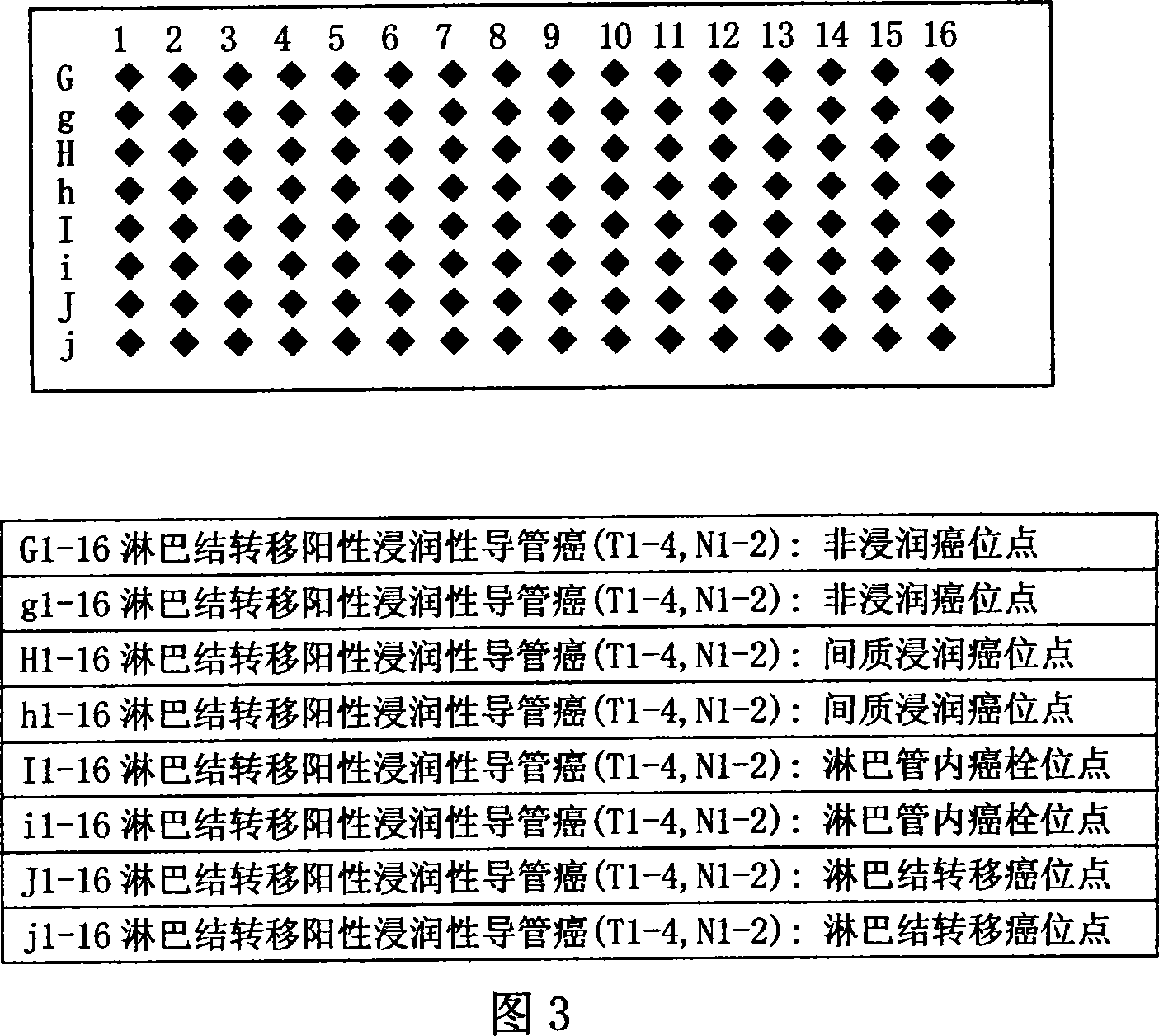
[0029] Example 1: Mimic tissue chip for breast ductal carcinoma lymphatic metastasis
[0030] (1) Collection of donor paraffin tissue specimens:
[0031] According to the WHO (2003) new histological classification of breast tumors, cases of minimally invasive ductal carcinoma and invasive ductal carcinoma (including positive and negative lymph node metastasis) were selected, and surgical resection specimens were collected in recent years.
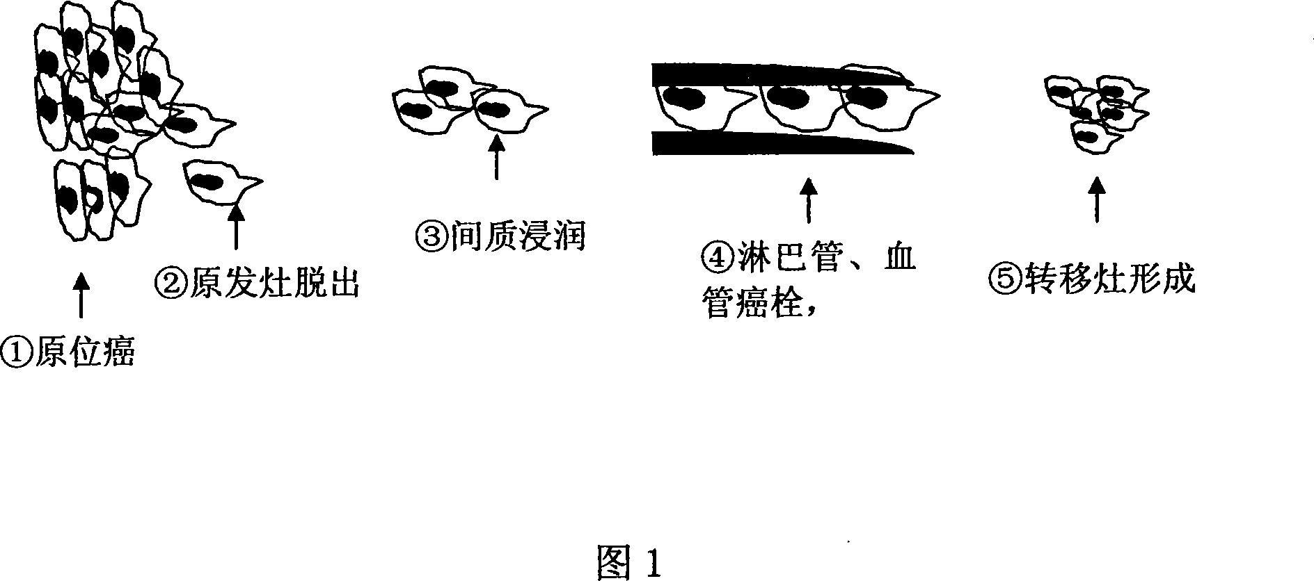
[0032] The basis for selecting microinvasive carcinoma and invasive ductal carcinoma (including positive and negative lymph node metastasis): ductal carcinoma of the breast occurs in the duct of the breast, and is confined to the duct in the early stage to form ductal carcinoma in situ. As the disease progresses, cells with invasive ability proliferate And break through the basement membrane to form micro-invasive carcinoma, and then, the infiltrating cancer cells infiltrate in the loose and edematous interstitium under the external interac...
Embodiment 2
[0047] Example 2: Colorectal Adenocarcinoma Blood Tract Metastasis Tissue Chip
[0048] (1) Collection of donor paraffin tissue specimens:
[0049] According to WHO (2000) colorectal tumor histology and TNM classification criteria, collected in recent years Tis (carcinoma in situ: intraepithelial or infiltrating lamina propria), T1 (tumor infiltrating submucosa), T2 (tumor infiltrating muscle layer), T3 ( Surgical resection of adenocarcinoma cases (including positive and negative liver metastases) to select paracancerous tissues, cancer tissues with different depths of invasion (invasive cancer and intravascular tumor thrombus), and liver metastases wax block specimen.
[0050] (2) Perform immunohistochemical staining on the above-mentioned donor paraffin tissue specimens:
[0051] T1, T2, and T3 specimens were serially sectioned, and conventional HE staining and lymphatic staining (such as vascular endothelial growth factor receptor-3, vascular endothelial growth factor rec...
Embodiment 3
[0062] Example 3: Gastric cancer tumor implantation and metastasis tissue chip
[0063] (1) Collection of donor paraffin tissue specimens:
[0064] According to the WHO (2000) gastric tumor histology and TNM classification criteria, Tis (carcinoma in situ: confined to the epithelial layer), T1 (tumor infiltrating the lamina propria or submucosa), T2 (tumor infiltrating the muscular layer), and T3 in recent years were collected. (Tumor infiltrating the subserosa without invading adjacent structures) and T4 adenocarcinoma with implantation and metastasis were surgically resected to select paracancerous tissues, tumor tissues infiltrating at different depths, and implanted and metastatic cancer tissues.
[0065] (2) Perform immunohistochemical staining on the above-mentioned donor paraffin tissue specimens:
[0066] T1, T2, T3, and T4 specimens were serially sectioned, and conventional HE staining and lymphatic staining (such as vascular endothelial growth factor receptor-3, vas...
PUM
 Abstract
Description
Claims
Application Information