

Real-time list mode reconstruction
By monitoring the position of the object support surface and recording the gamma ray reception time, the data in the PET scanner is analyzed and reconstructed in real time, which solves the problem of delay in the PET scanner data acquisition and reconstruction process, achieves rapid image reconstruction, and increases processing throughput.
- Summary
- Abstract
- Description
- Claims
- Application Information
AI Technical Summary
Problems solved by technology
Method used
Image

Examples
Embodiment Construction
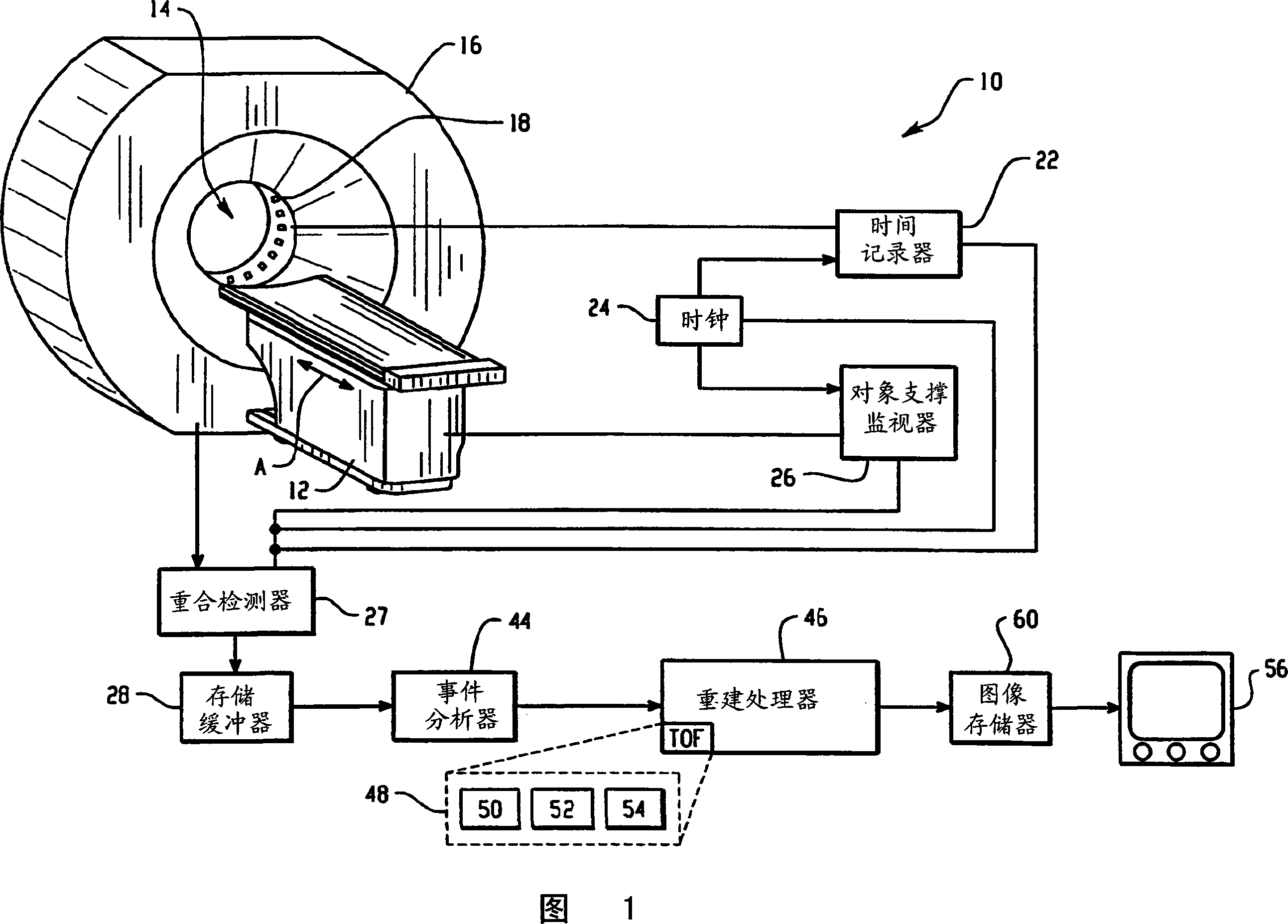
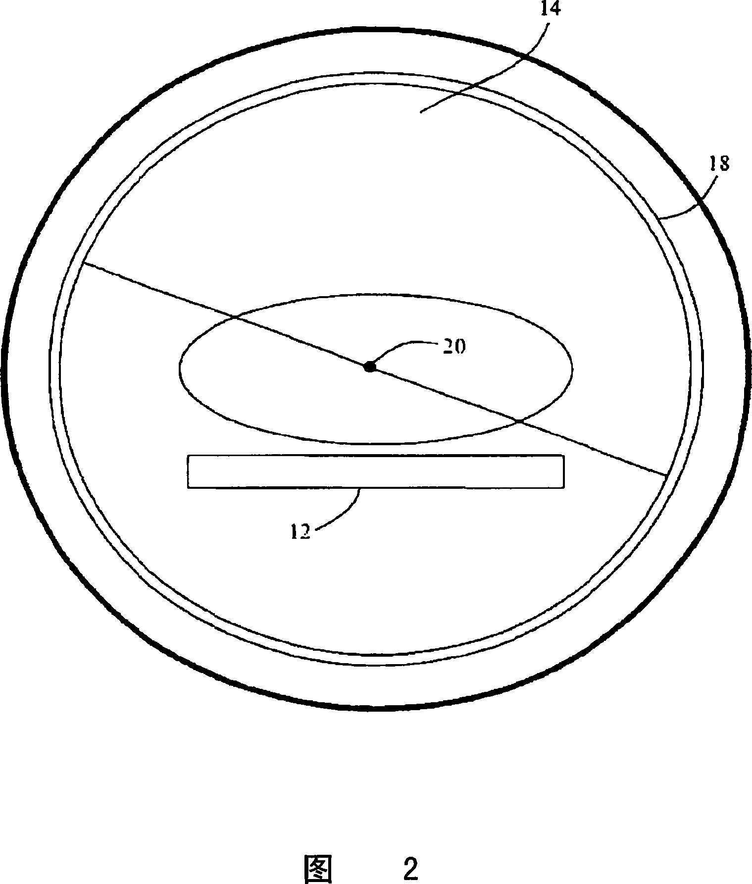
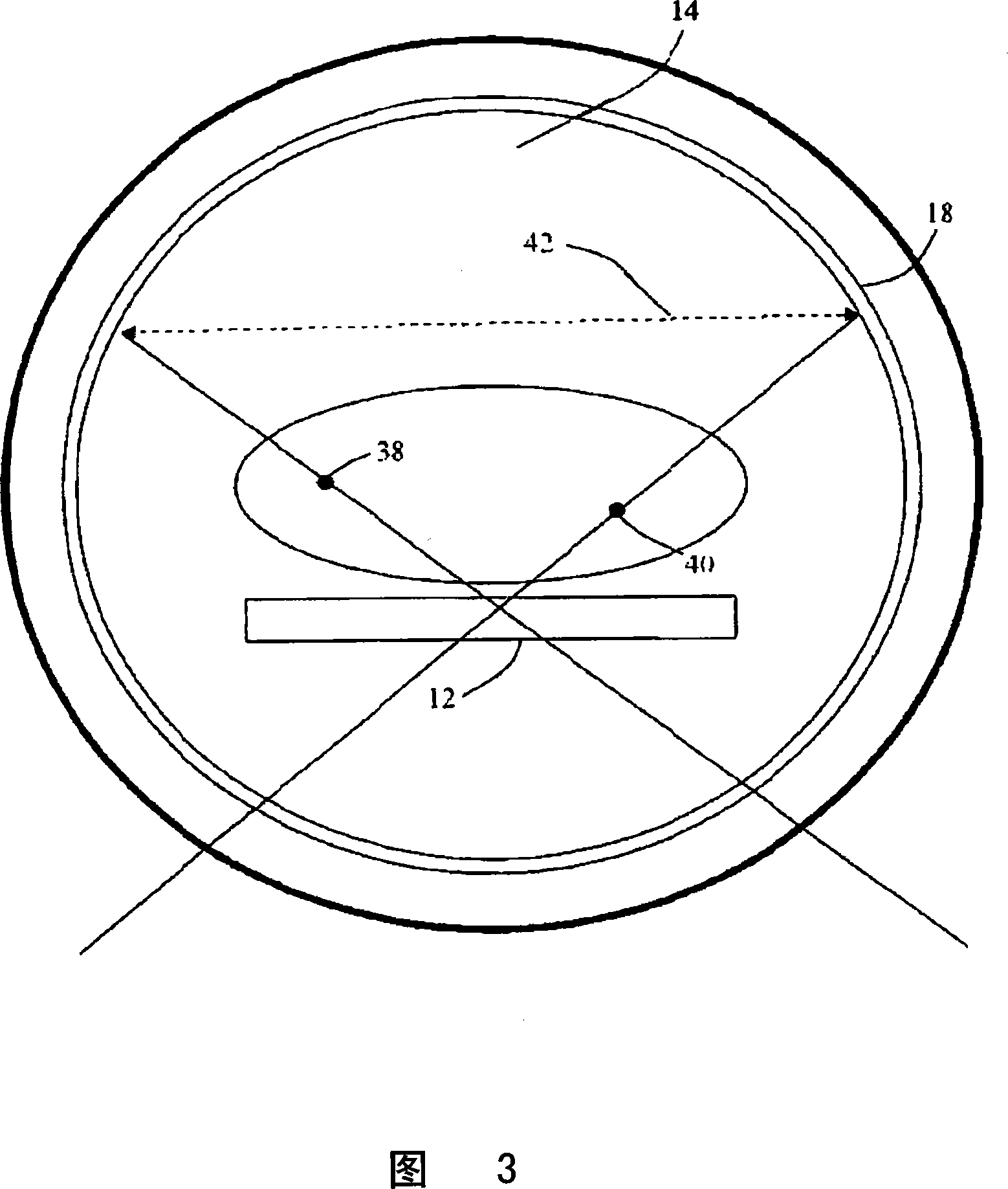
[0015] Referring to Figure 1, a preferred embodiment of a nuclear medicine scanner 10 is shown. The object is placed on the object support surface 12 prior to scanning. The object support surface moves along its longitudinal axis A into and out of the cavity 14 of the gantry 16 of the scanner 10 . The lumen of the PET scanner is lined with the cylinder of the radiation detector 18 . Optionally, the detector comprises a plurality of detection heads. In either case, detectors 18 are positioned around and along the subject receiving lumen 14 to receive nearly simultaneously incident gamma rays. Typically, incident gamma rays impinge on a detector 18, which preferably includes a scintillation crystal and a photodetector array, although solid state, Anger-type and other detectors are also contemplated. When the scintillation crystals are struck by gamma rays, they emit small pulses of visible light, which are detected by photodetectors and converted into electrical signals. A s...
PUM
 Abstract
Description
Claims
Application Information