Protein delivery system

A technology of protein and protein fusion, which is applied in the field of delivery of protein substances to cells, and can solve problems such as the difficulty of delivering proteins to cells

Inactive Publication Date: 2008-03-19
BIOACTIVE PROTEIN DELIVERY THERAPEUTICS
View PDF2 Cites 5 Cited by
  • Summary
  • Abstract
  • Description
  • Claims
  • Application Information

AI Technical Summary

Problems solved by technology

[0010] While hopeful, delivering protein in sufficient quantities to cells has been elusive

Method used


Image

  • Protein delivery system
  • Protein delivery system
  • Protein delivery system

Examples


Embodiment 1

[0134] Example 1: Fabrication of CFTR-GFP VLPs

[0135] A chimeric gene encoding green fluorescent protein (GFP) fused to CFTR with a GFP domain at the N-terminus of the CFTR protein was incorporated into the VLPs. The GFP moiety-tagged CFTR enables rapid recognition and enables rapid establishment of the tagged protein. The fusion gene GFP-CFTR was cloned in two different expression systems, (i) baculovirus AcMNPV to form recombinant baculovirus AcMNPV-GFPCFTR, (ii) in adenovirus type 5 (Ad 5) to produce recombinant virus Ad 5-GFP-CFTR. The protein was observed to localize to the plasma membrane of the cell. Similar GFP-tagged structures carried by expression plasmids have been reported to have the same function as the non-tagged protein CFTR (Haggie et al., 2002, J. Biol. Chem. 277:16419-16425 and Loffing-Cueni et al., 2001, Am. J .Physiol.Cell Physiol.281:C1889-1897.20).GFP-CFTR clones were shown to be activated and functional (Robert et al., 2004, J.Biol.Chem.279:21160-...

Embodiment 2

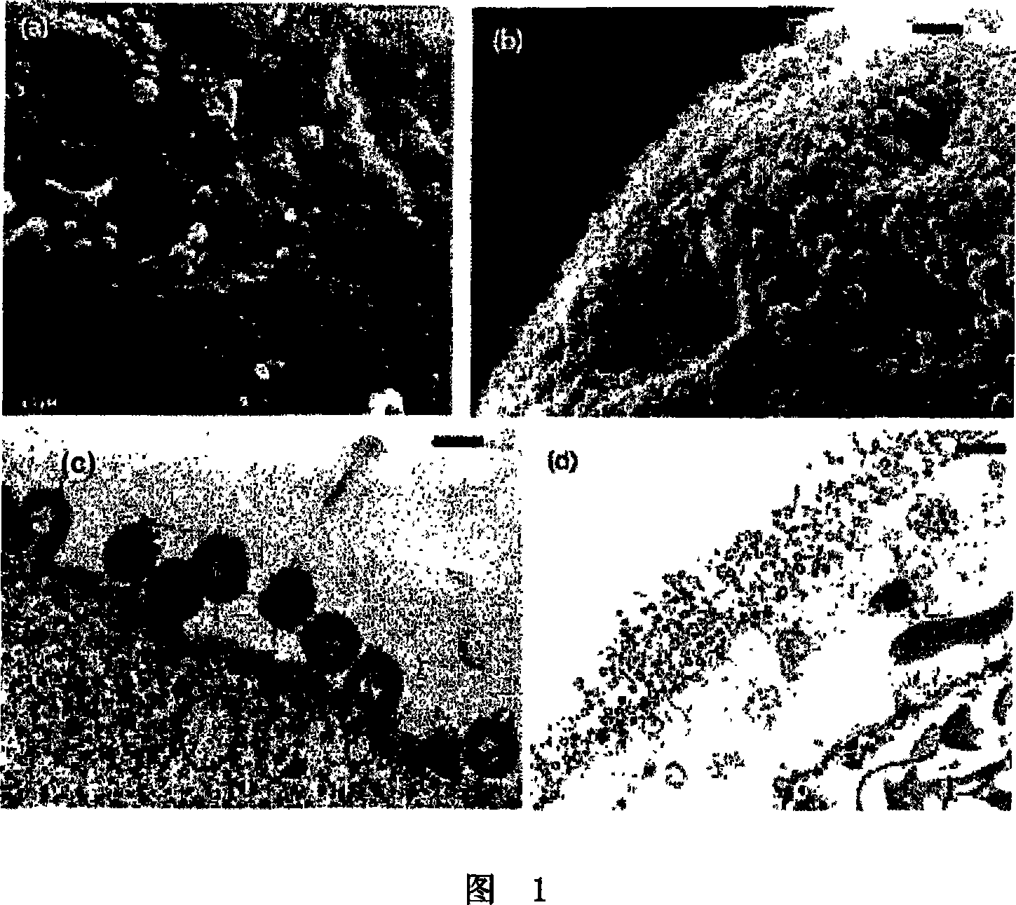
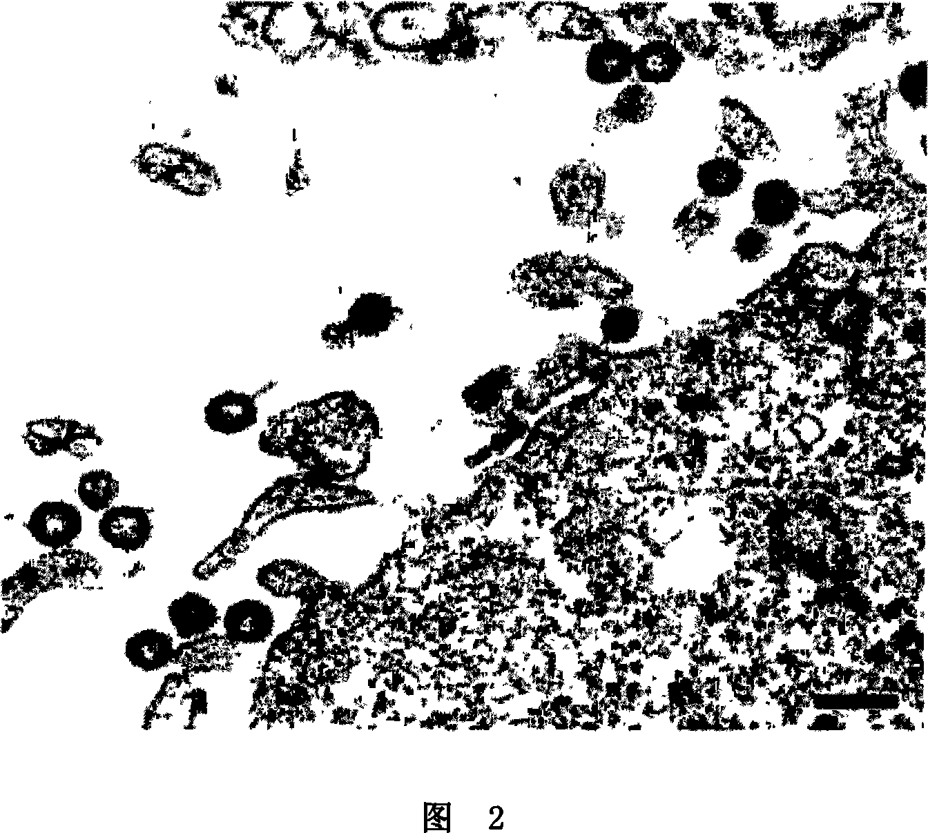
[0140] Example 2: Transmission Electron Microscopy

[0141] Cells were infected with the indicated viruses at a multiplicity of infection (MO1) of 25 as indicated in the legend. Cells were collected for electron microscopy (EM) examination by pelleting by centrifugation 48 hours post-infection. Cell pellets were then fixed with 2.5% glutaraldehyde in 0.1 mpH 7.5 phosphate buffer, post-fixed with osmium tetroxicle, and treated with tannic acid (0.5% in H2O). After dehydration, samples were embedded in epoxy resin (Epon) (Epon-812, Fulham, Latham, New York). Sections were extracted, stained with 2.6% basic lead citrate-0.5% Sections uranyl acetate in 50% ethanol, and examined under a Jeol 1200-EX electron microscope.

Embodiment 3

[0142] Example 3: Scanning Electron Microscopy

[0143] Cells were grown on glass slides and infected at a multiplicity of infection (MOI) of 25 with the viruses indicated. Fixation and postfixation were the same as described for transmission EM. Cells were then point dried with liquid CO2, covered with gold, and examined with a Zeiss DSM 950 electron microscope.


PUM


Abstract

The present invention relates to a virus-like particle (VLP) having a plasma membrane-derived lipid bilayer envelope, said VLP further comprising a viral structural protein, or fragment or derivative thereof, capable of forming an enveloped VLP, a fusiogenic protein and a recombinant target protein; methods for the delivery of recombinant target proteins to cells using said VLP, therapeutic methods using said VLP, compositions and kits comprising said VLP, methods of producing said VLP, and vectors and host cells for producing said VLP are also described.

Description

technical field [0001] The present invention relates to the delivery of non-genetic material to cells, in particular to the delivery of protein material to cells. Background technique [0002] Currently, gene therapy for inherited or acquired diseases, such as cystic fibrosis or cancer, generally involves the delivery of one of two therapeutic sequences. The first method uses naked nucleic acid or non-viral vectors, which are usually encapsulated liposomes or lipoplexes. The second method uses viral vectors. Viral vectors can be non-conjugated, such as adenovirus (Ad) or herpes virus (HSV); viral vectors can also be conjugated, such as adeno-associated virus (AAV) and denatured virus (eg MLV). In the case of Ad and HSV, expression of the therapeutic gene is only transient. In the case of bound vectors, such as denatured viruses or AAV, expression is long-term (theoretical cell life span). [0003] These gene therapy approaches have several disadvantages. The delivery ef...

Claims


Application Information

Patent Timeline
Patent Type & Authority Applications(China)
IPC IPC(8): A61K9/50
CPCA61K47/48776C12N2760/20222C12N7/00C12N2740/16122C12N2740/16023C07K14/005A61K38/00A61K9/5184A61K47/6901A61P1/18A61P11/00A61P35/00Y02A50/30
Inventor 斯帝恩·林德卡尔·詹森
Owner BIOACTIVE PROTEIN DELIVERY THERAPEUTICS
Why Eureka
Try Eureka