Portable sample rapid joint inspection device

A portable, sample technology, used in material testing products, instruments, and analysis by chemical reaction of materials, etc., can solve the problems of insurmountable cross-interference, time-consuming separation of serum, and limited scope of application, so as to avoid detection cross-interference. , to avoid the spread of pathogens, and to broaden the scope of application

Active Publication Date: 2011-04-13
马义才
View PDF7 Cites 0 Cited by
  • Summary
  • Abstract
  • Description
  • Claims
  • Application Information

AI Technical Summary

Problems solved by technology

[0003] Chinese patent ZL.200420096377.3 and application number 200410080305.4 disclose a rapid diagnostic test tube for vacuum blood collection. Although the device can be used for joint inspection of several samples, it can also better solve the problems of iatrogenic infection and environmental pollution, but it has disadvantages Yes: (1) The structure of the device is complicated, the manufacturing cost is high, and it is difficult to popularize
(2) The scope of application is limited
The device is only suitable for on-the-spot blood collection and detection by health technicians who have undergone certain training, but it is powerless for some samples that have been collected and prepared, especially for the detection of non-blood samples that have been collected and prepared, such as Patient urine, separated blood, plasma and serum, saliva, body fluids, secretions and other liquid samples (such as liquid samples of drugs, food, pesticide residues) that have been submitted or collected for preparation, etc.
(3) The use of vacuum test tubes for blood collection is far from being popular in grassroots institutions and rural areas in my country, and sometimes iatrogenic infections, pathogens spread, and environmental pollution have occurred due to failure of blood collection. For those who have not been specially trained For ordinary people, its use is more difficult
In addition, Chinese patent application No. 200410099301.0 discloses a colloidal gold test strip that can jointly detect multiple pathogens, and Chinese patent application Nos. 02112230. Although these colloidal gold test strips and colloidal gold percolation devices can achieve various indicators in the joint inspection samples, there are still deficiencies: (1) the joint inspection indicators are all placed on a test paper, and each detection The cross-interference existing between
(2) Blood samples need to be separated from serum first, not suitable for detection of whole blood or other viscous samples
Moreover, separating serum is not only time-consuming, but also requires certain equipment, reagents and conditions

Method used


Image

  • Portable sample rapid joint inspection device
  • Portable sample rapid joint inspection device
  • Portable sample rapid joint inspection device

Examples


Embodiment 1

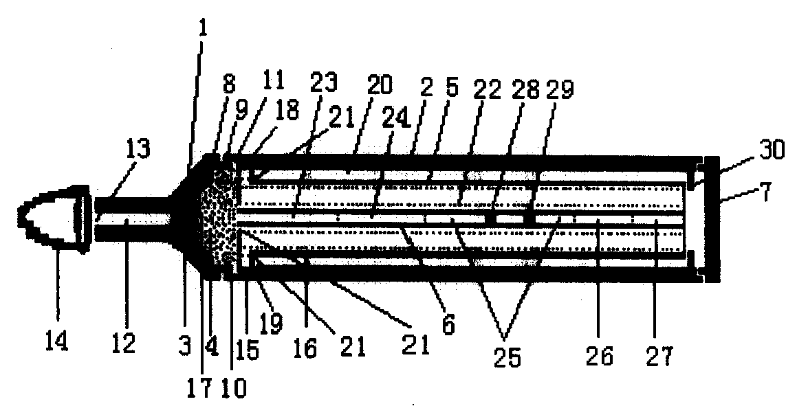
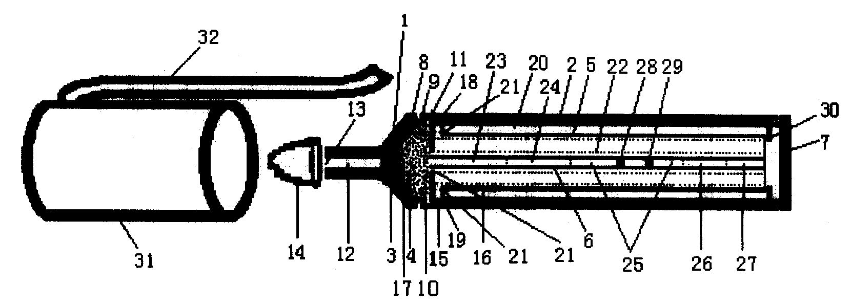
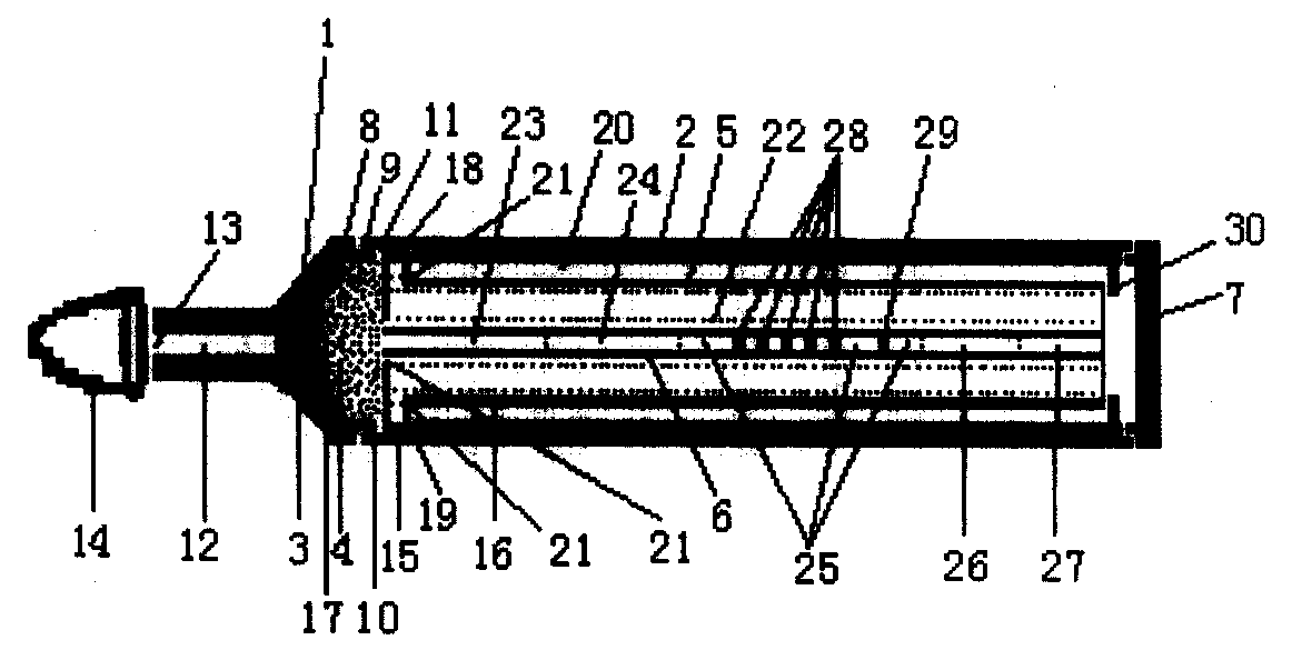
[0042] Embodiment 1 The structure of the portable sample rapid joint inspection device (illustrated in conjunction with the accompanying drawings)

[0043] figure 1 It is a side view of the structure of the rapid joint inspection device for samples composed of built-in single detection line test strips of the present invention. The device includes a sample suction head 1 and a detection tube 2 connected to the back end of the sample suction head 1 .

[0044] A capillary suction nozzle 12 is arranged at the front end of the suction head 1 . The suction nozzle 12 is provided with a sample suction port 13 for sample suction. The front end of the suction nozzle 12 is covered with a rubber protective cap 14. During testing, the rubber protective cap 14 is removed to expose the sample suction port 13, and the joint inspection of samples can be performed by dipping the suction nozzle 12 in the inspection product. The cavity 17 of the suction head is filled with sample filter mate...

Embodiment 2

[0057] Example 2 Operation method of portable sample rapid combined inspection device

[0058] The sample to be tested applicable to the joint detection device of the present invention can be from clinical or non-clinical blood (including whole blood, serum, plasma), body fluid, urine, saliva, reproductive tract secretion or other liquid samples or viscous samples. sample. Among them, clinical samples include samples of infectious diseases, hormones, cardiovascular diseases, tumors, autoimmune diseases, drugs, etc., non-clinical samples include food testing, environmental pollution testing, biological contamination testing, biological agent testing, veterinary testing, Samples for forensic testing, etc.

[0059] The operation method of the joint inspection device is as follows:

[0060] (1) Take out the sample rapid joint inspection device;

[0061] (2) Remove the device head cover 31 of the device and the rubber cap 14 at the front end of the suction head to expose the sam...


PUM


Abstract

The invention discloses a portable and fast sample joint inspection device consisting of a sample suction head (1) and a test tube (2) connected with the back end of the sample suction head (1), wherein, the front end of the sample suction head (1) is provided with a tubule suction nozzle (12) provided with a suction port (13) used for sucking samples; the front end of the suction nozzle (12) canbe sheathed with a rubber protecting cap (14); the cavity(17) of the suction head is respectively filled from forth to back with the sample of filter material (3) and the sample of water absorbent material (4); a fixation column of test paper (5) is arranged in the test tube (2); one to a plurality of sample test paper strips (6) are fixed on the fixation column of test paper (5); the tail end ofthe test tube is provided with a seal cover (7) for enclosing the tested waste in the test tube (2) without leakage. When each sample test paper strip (6) enclosed and fixed in the device goes through sample suction for once and the same background detection, a plurality of indexes of the samples can be jointly detected at the same time by naked eyes in a plurality of minutes, therefore, a plurality of sample tests become simpler, safer and faster, and can avoid cross interference. The tested waste remains sealed in the test tube to be more beneficial to avoiding pathogen spreading and iatrogenic infection as well as treating waste materials in an environment protection way.

Description

technical field [0001] The invention relates to a test paper for joint inspection of medical equipment and samples, in particular to a portable rapid joint inspection device for samples. Background technique [0002] Colloidal gold immunochromatography (Gold-Immunochromatography Assay, GICA) is an immunolabeling technique developed on the basis of enzyme-linked immunosorbent assay (ELISA). The capillary action of the membrane makes the liquid sample dropped on one end of the membrane strip slowly seep to the other end of the microporous membrane. If there are specific antigens or antibodies in the liquid, they can be combined with immunogold and then fixed on the microporous filter. The corresponding antibody or antigen on the membrane binds to show the immunogold color. This technology is simple, fast, and does not require any equipment, and the results can be observed with the naked eye in a few minutes. It is known as "concentrated ELISA". Colloidal gold immunochromatog...

Claims


Application Information

Patent Timeline
Patent Type & Authority Patents(China)
IPC IPC(8): G01N21/78G01N33/52G01N35/10
Inventor 马义才顾敏马灵
Owner 马义才
Why Eureka
Try Eureka