
Method for detecting identical antigen expression on identical sample

A technique for specimens and antigens, applied in the field of immunopathology detection, can solve the problems of inconsistent experimental conditions, labor-intensive, material resources, and biased results, and achieve the effect of less harsh experimental conditions, low background, and consistent conditions

Inactive Publication Date: 2008-10-29
WUHAN UNIV
View PDF0 Cites 11 Cited by
  • Summary
  • Abstract
  • Description
  • Claims
  • Application Information

AI Technical Summary

Problems solved by technology

[0007] At present, almost all literature reports using immunofluorescence technology and immunoenzyme technology to detect antigen expression are carried out on two specimens separately, which will make the experimental conditions inconsistent, and will also make the antigens detected by these two technologies separately The results are biased
At the same time, it consumes more manpower and material resources
However, so far, there is no report similar to our use of immunofluorescence technology and immunoenzyme technology on the same specimen to detect the expression of the same antigen

Method used

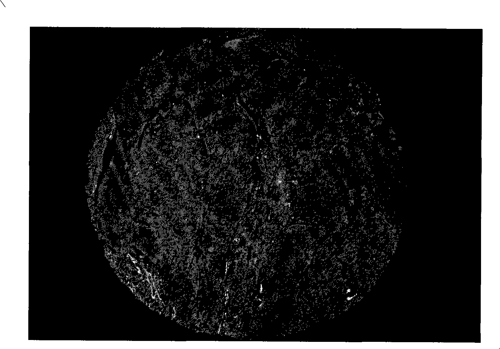

Image

  • Method for detecting identical antigen expression on identical sample
  • Method for detecting identical antigen expression on identical sample
  • Method for detecting identical antigen expression on identical sample

Examples


example 1

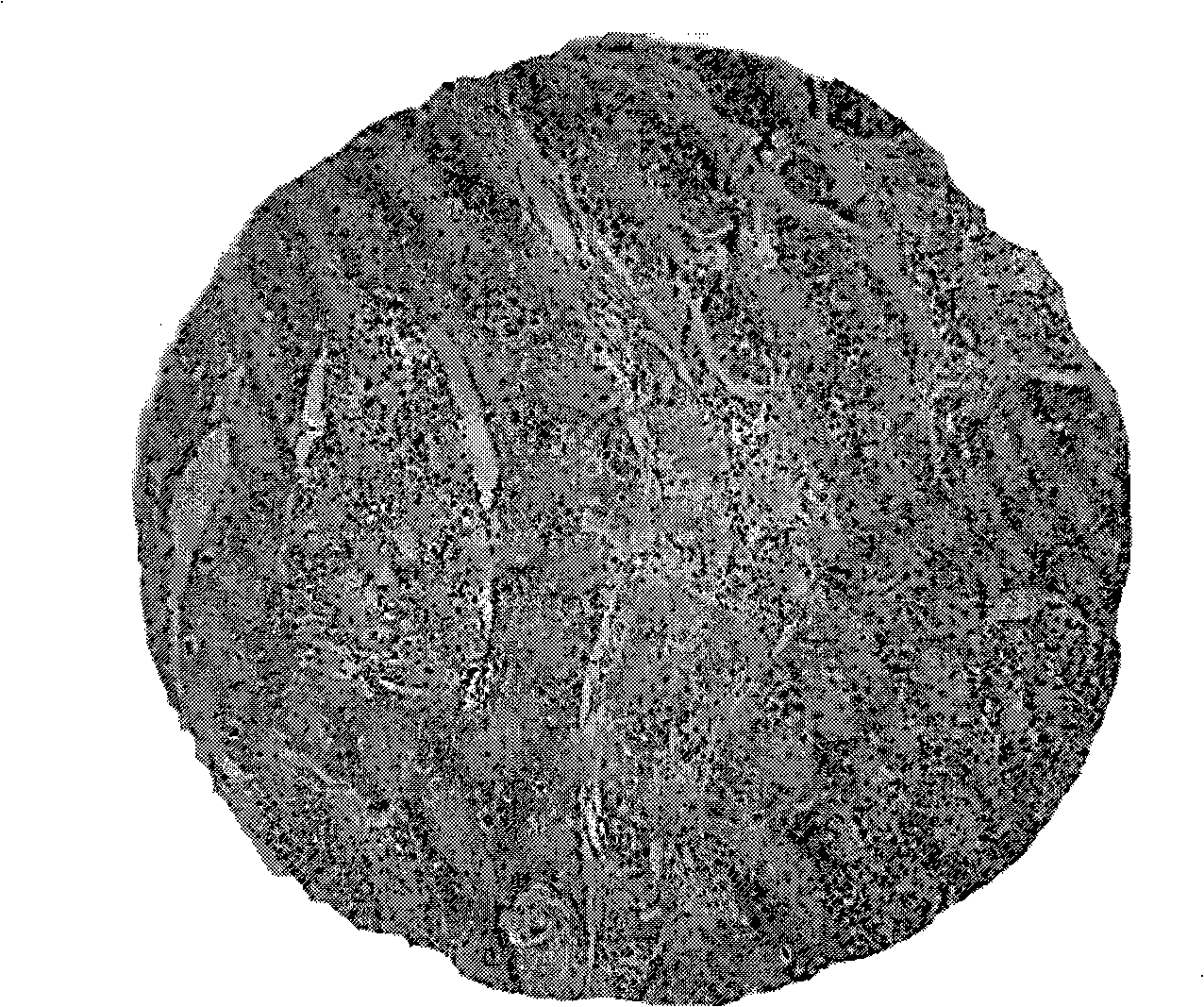
[0030] Example 1 detects the expression of Caveolin-1 protein on a lung cancer tissue chip, and the steps are:

[0031] 1.4 μm thick lung cancer tissue chips were dewaxed, hydrated, and washed with TBS (pH7.4) for 2 to 4 times, each time for 3 minutes;

[0032] 2. 0.01M pH6.0 citrate buffer, microwave antigen retrieval, naturally cool to room temperature (20-25°C, the same below), wash with TBS 2-4 times, 3min each time;

[0033] 3.2% BSA (bovine serum albumin) blocking buffer (that is, 2g BSA dissolved in 100ml TBS solution) and incubated in a wet box at 37°C for 30-35min;

[0034] 4. Add Caveolin-1 (1:100) antibody dropwise, incubate in a wet box at 37°C for 1.5-2 hours or in a refrigerator at 4°C overnight, rinse with TBS-T (pH7.4) for 3min / time x (2-4) times;

[0035] Incubate with 5.2% BSA blocking buffer in a wet box at 37°C for 10-15 minutes;

[0036] 6. Add biotinylated goat anti-rabbit IgG dropwise, incubate in a wet box at 37°C for 30-35 minutes, rinse with TBS-T f...

example 2

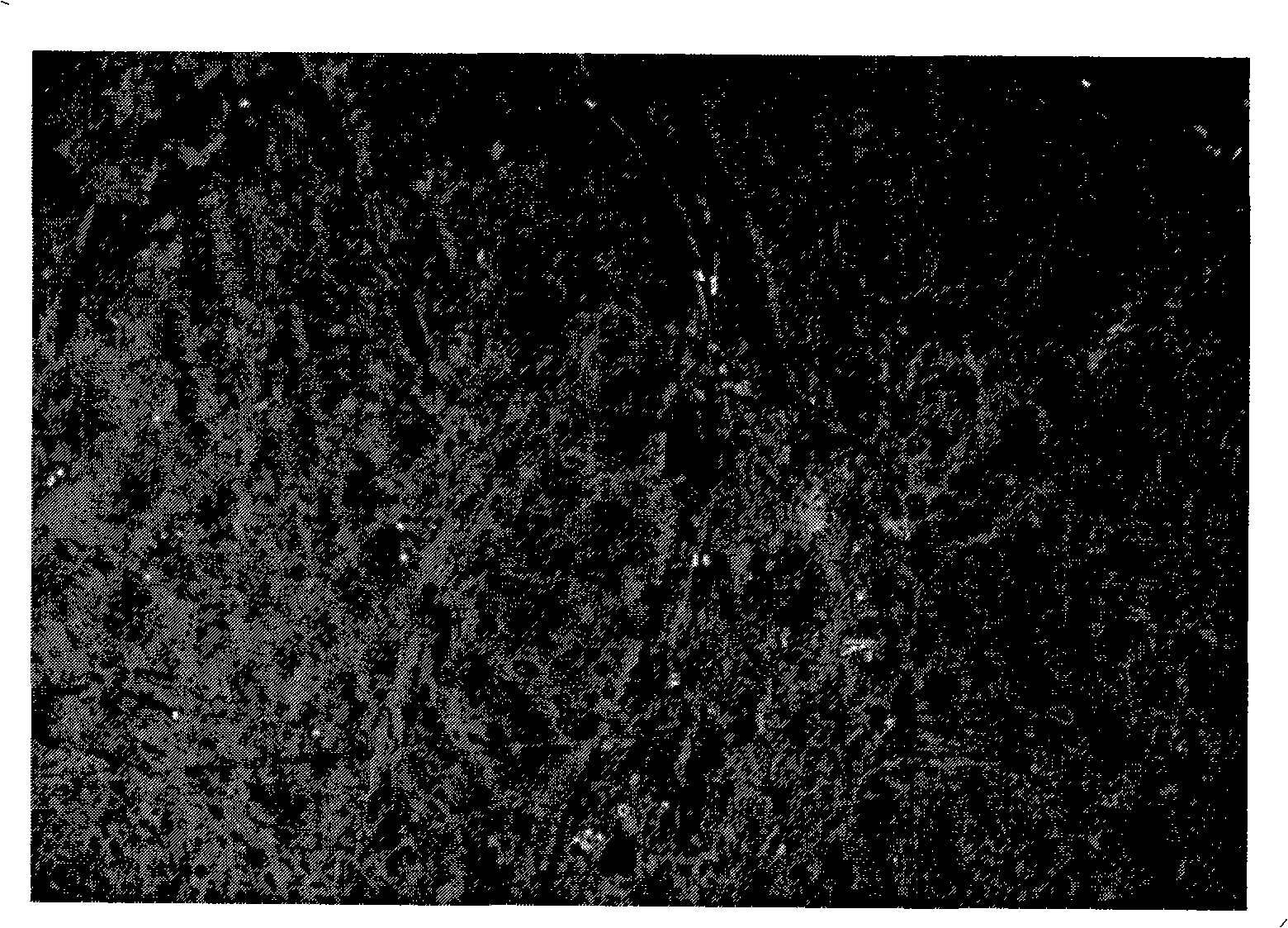
[0045] Example 2 detects the expression of Caveolin-1 protein on lung cancer cell line A549 cells, and the steps are:

[0046] 1. After A549 cells are fixed, add TBS (pH7.4) to wash 2-4 times, each time for 3 minutes, then add permeabilization solution (0.1% Triton-X 100) and incubate at room temperature for 5-10 minutes, and finally wash with TBS 2-4 times , 3 minutes each time;

[0047] Incubate with 2.2% BSA blocking buffer in a humid box at 37°C for 30-35 minutes;

[0048] 3. Add Caveolin-1 (1:100) antibody dropwise, incubate at 37°C for 1.5-2 hours or overnight in a refrigerator at 4°C, rinse with TBS-T (pH7.4) for 3min / time x (2-4) times;

[0049] Incubate with 4.2% BSA blocking buffer in a wet box at 37°C for 10-15 minutes;

[0050] 5. Add biotinylated goat anti-rabbit IgG (50-200μl) dropwise, incubate in a wet box at 37°C for 40-45min, rinse with TBS-T for 3min / time x (2-4) times;

[0051] Incubate with 6.2% BSA blocking buffer in a humid box at 37°C for 10-20 minut...


PUM


Abstract

The invention discloses a method for detecting the same antigen expression on the same specimen. A fluorescent quantum dot marked streptavidin compound can be directly combined with a biotin conjugate second antibody, while the biotin conjugate second antibody can also be combined with horseradish peroxidase marked streptavidin in immunoenzyme technique, so that the immunofluorescence and immunoenzyme techniques can be perfectly combined, thereby detecting the same antigen expression on the same specimen. The method can be used for detecting the antigen expression state of the fresh organizations of human bodies and animals, paraffin embedding organizations, and the states of antigen expression in different cell lines. The method has the advantages of higher sensitivity, more persuasive results, low experiment cost, and moderate requirements for experimental conditions. The whole experiment process does not need to avoid light, and can be completed in an ordinary immunohistochemical room. The method is suitable for popularization and application.

Description

technical field [0001] The invention belongs to the field of immunopathology detection, and more specifically relates to a method for detecting the expression of the same antigen on the same specimen using two techniques, and the scope of application of the method is suitable for detecting fresh tissues, paraffin-embedded tissues and different cell lines of humans and animals Antigen expression within. Background technique [0002] Immunohistochemistry, also known as immunocytochemistry, is the basic principle of applied immunology - antigen-antibody reaction, that is, specific antibodies labeled with chromogenic agents (fluorescein, enzymes, metal ions, isotopes) It is a technology for qualitative, localization and quantitative determination of the corresponding antigen through antigen-antibody reaction and histochemical color reaction in situ on tissue cells. It skillfully combines the specificity of the immune response and the visibility of histochemistry, and detects va...

Claims


Application Information

Patent Timeline
IPC IPC(8): G01N21/64G01N21/76G01N1/28
Inventor 陈洪雷高俊张玉霞余保平
Owner WUHAN UNIV
Learn More