
Novel method for quantitatively determining analyte by scavenger with single specificity

A quantitative detection method and analyte technology, applied in the field of bioengineering, can solve the problems of interference with specific analyte labeling efficiency, failure to distinguish from specific analytes or non-specific substances, and reduce antibody binding.

Inactive Publication Date: 2009-02-04
孙东旭
View PDF0 Cites 27 Cited by
  • Summary
  • Abstract
  • Description
  • Claims
  • Application Information

AI Technical Summary

Problems solved by technology

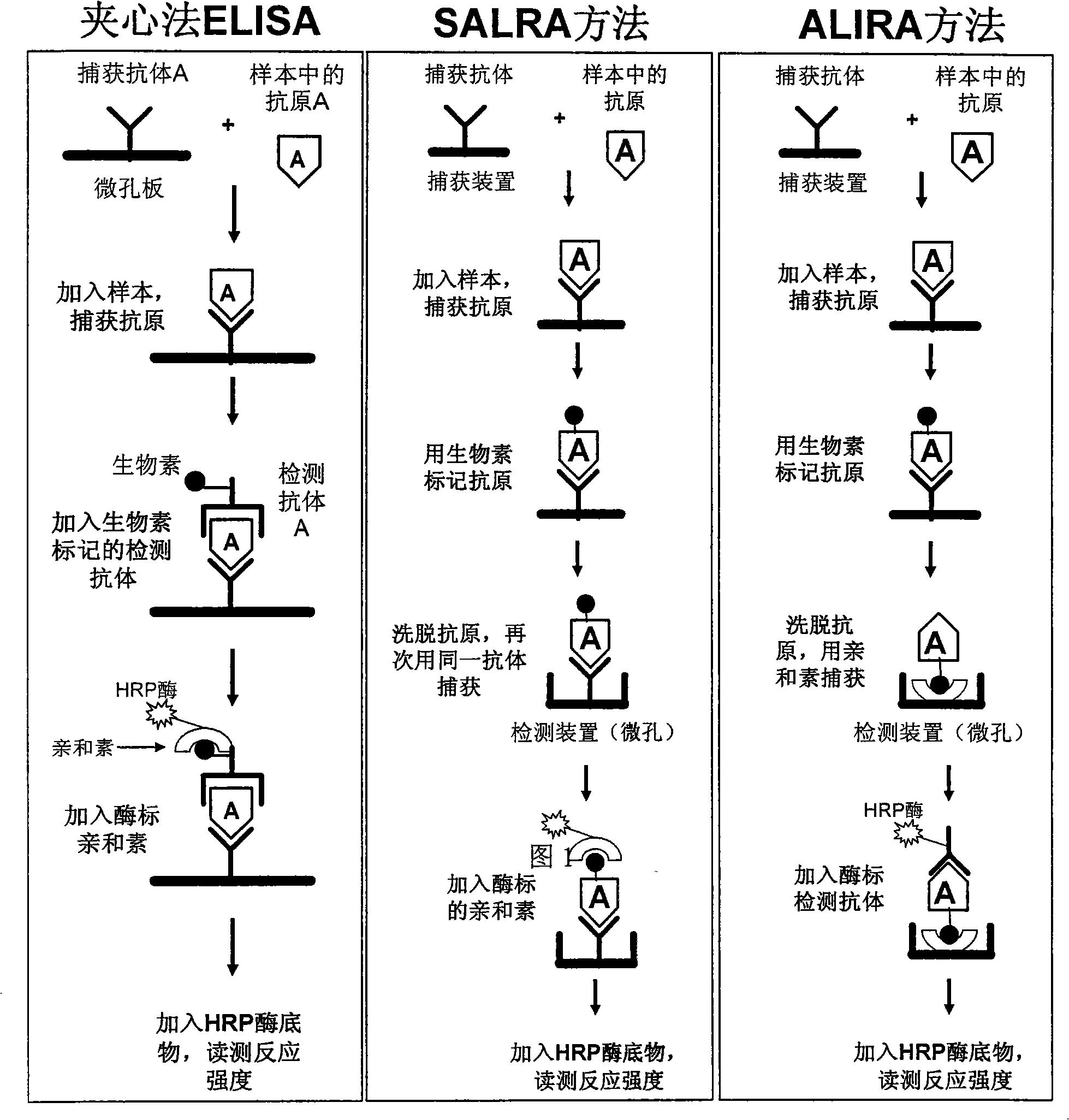
Although the specificity and sensitivity of the sandwich ELISA are relatively high, the application of this method has an important limitation: if you want to establish a sandwich ELISA detection method for the detection of an analyte, you must have two different antibodies against the analyte (capture antibody and detection antibody), and these two antibodies are required to have high specificity and high affinity for the analyte, and they must cooperate with each other, that is, the binding of the capture antibody and the analyte does not affect the detection antibody to the same Binding of analyte molecules
Although this method is simple and easy, and only needs one antibody (capture antibody) to detect, it has the following disadvantages: First, biological samples often contain thousands of molecules of various sizes, many of which will interfere directly or indirectly. Labeling efficiency for specific analytes, proteins at low concentrations tend to be more difficult to label efficiently
Second, the labeling process itself is likely to modify and change the epitope characteristics on the analyte molecule, reducing or even inhibiting the binding to the capture antibody on the solid phase.
Third, the overwhelming majority of non-specific substances in the sample have been effectively labeled before being cleared. Since the analyte has not been specifically enriched (concentrated), these non-specific substances that also carry reporter molecules will produce a strong The detection background noise greatly reduces the specificity and sensitivity, which has been proved by many experimental results using microwell plates or protein microarrays as detection platforms
Once bound to the surface of the detection device, the labeled molecules (reporter molecules) carried on these non-specific substances will generate background noise, and at this time the detection system has no steps to distinguish whether the reporter molecules are from specific analytes or from non-specific substances (mechanism)

Method used


Image

  • Novel method for quantitatively determining analyte by scavenger with single specificity
  • Novel method for quantitatively determining analyte by scavenger with single specificity
  • Novel method for quantitatively determining analyte by scavenger with single specificity

Examples


Embodiment 1

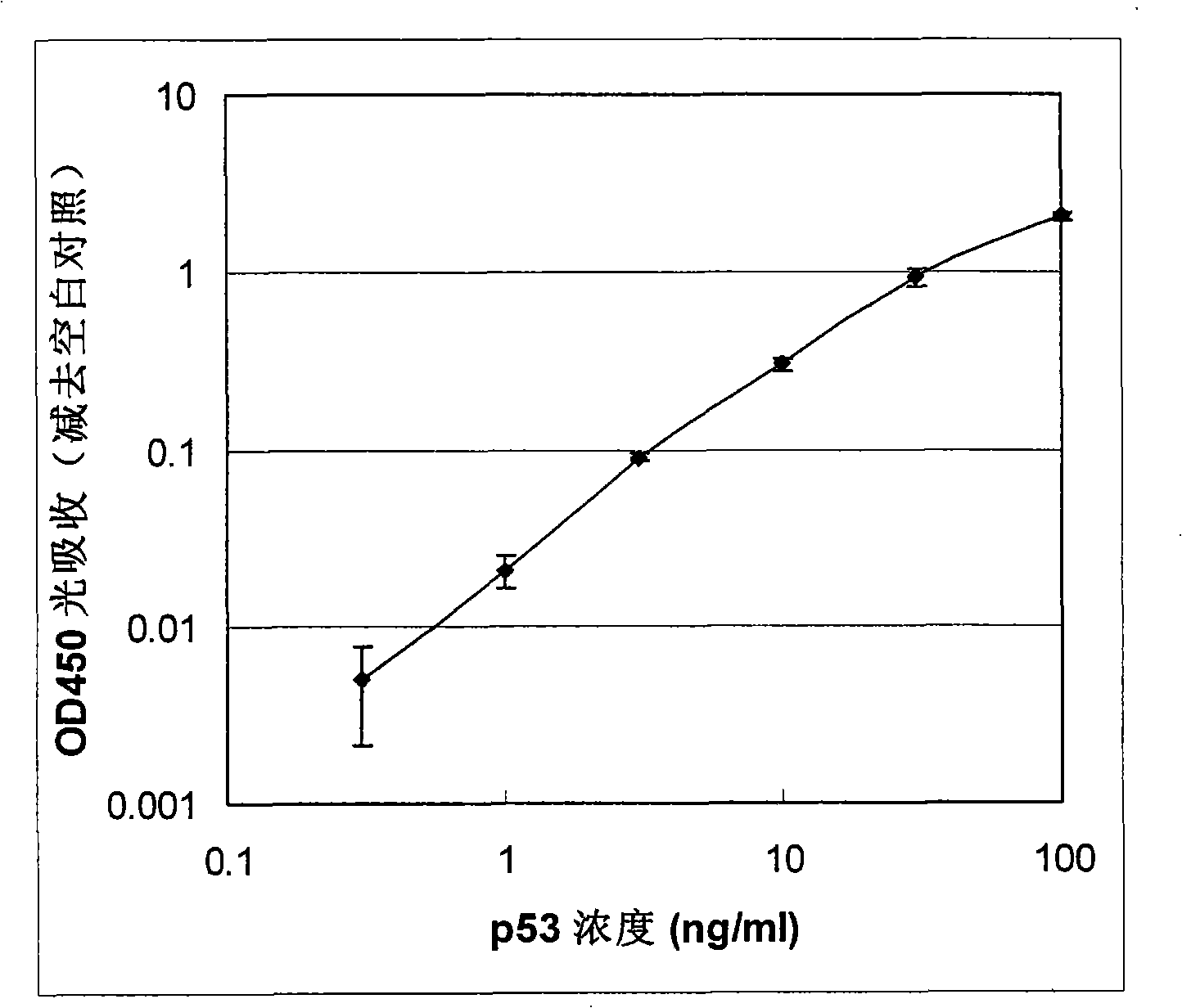
[0061] Embodiment 1: Use HRP as reporter molecule to detect human p53 protein

[0062] In this example, the antigen protein to be tested is human p53 protein which has tumor suppressive function and promotes cell apoptosis, and both the capture device and the detection device are 96-well microplates. The microwells of the capture device are coated with anti-p53 monoclonal antibody, and the detection device is coated with streptavidin. The detection antibody is the same anti-p53 monoclonal antibody labeled with fluorescein, here the reporter molecule is fluorescein.

[0063] (1) Capture antigenic protein:

[0064] a. Coating capture antibody: Take two 96-well microplates (flat bottom, highly binding to protein), one is used as a capture device to capture the antigen in the sample, add 100 μl concentration of 2 μg / ml (diluted in Coating buffer, 0.05M sodium bicarbonate buffer, pH 9.6) anti-p53 monoclonal antibody (CalBiochem, USA), was added to two longitudinal rows of 16 micr...

Embodiment 2

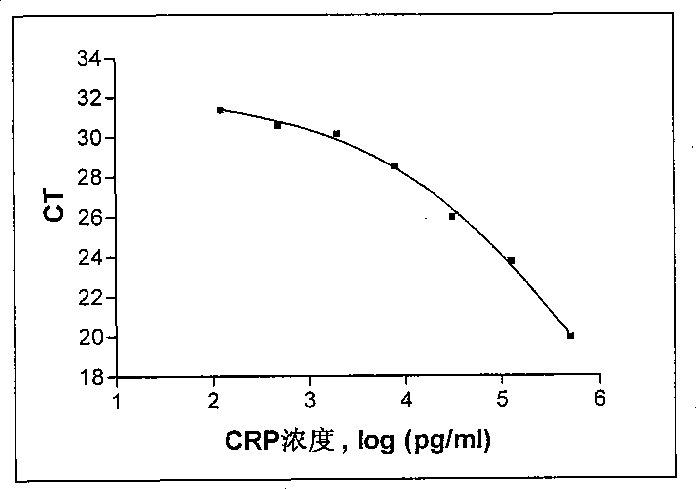
[0077] Example 2: Detection of human C-reactive protein with oligonucleotides as reporter

[0078] In this embodiment, the antigenic protein to be tested is human C-reactive protein (C-reactive protein, CRP), which plays an important role in the immune response, and both the capture device and the detection device are 96-well microplates. The microwells of the capture device are coated with anti-CRP monoclonal antibody, and the detection device is coated with streptavidin. The detection antibody was the same anti-CRP monoclonal antibody labeled with an oligonucleotide (60 bases long), where the reporter molecule was the oligonucleotide.

[0079] (1) Capture antigenic protein:

[0080] a. Coating capture antibody: Take two 96-well microplates (flat bottom, highly binding to protein), one is used as a capture device to capture the antigen in the sample, add 100 μl concentration of 2 μg / ml (diluted in Coating buffer, 0.05M sodium bicarbonate buffer, pH 9.6) anti-CRP monoclonal ...


PUM


Abstract

The present invention discloses a method which adopts a single specific capture agent for quantitative detection of the analyte in the biological sample, which is named as the analyte labeling and inversion recapture method, ALIRA for short, which is an improved detection method which is based on the principles of the SALRA but adopts different capture means. The method comprises: capturing the analyte on the capture device, and forming capture agent-analyte complex; labeling the complex with a labeling agent; eluting the labeled analyte from the complex; recapturing the analyte through the combination of the second recapture agent on the detection device and the labeling molecules; and adding specific detection agent, compounding the specific detection agent and the analyte, and determining the concentration of the analyte according through detecting the signals which are generated by the report modules which are carried by the specific detection agent. The invention has the advantage that the adoption of a specific detection agent can sensitively and simply complete the quantitative detection of the analyte, and the invention has excellent application prospects in the application in the disease diagnosis, medical evaluation, new drug development, protein micro-array and the like, and in the field of basic research.

Description

technical field [0001] The invention belongs to the technical field of bioengineering, and specifically relates to a new method for quantitatively detecting an analyte using a specific capture agent, which is applicable to various detection platforms based on solid phase surfaces, including microporous plates, filter membranes, micro Magnetic balls, and more. Background technique [0002] Detecting the content of specific protein factors in biological (including human) tissue samples is of great significance for basic research and applications in the fields of medicine, biology, agriculture, food industry, and environmental protection. For example, the detection of specific protein components of pathogenic microorganisms is currently the main means of diagnosing infectious diseases; similarly, the determination of changes in the levels of early biomarker protein factors for cancer and cardiovascular diseases is extremely important for early detection, early treatment and cur...

Claims


Application Information

Patent Timeline
Patent Type & Authority Applications(China)
IPC IPC(8): G01N33/543G01N33/68G01N33/577
Inventor 孙东旭斯蒂芬·诺克
Owner 孙东旭
Learn More