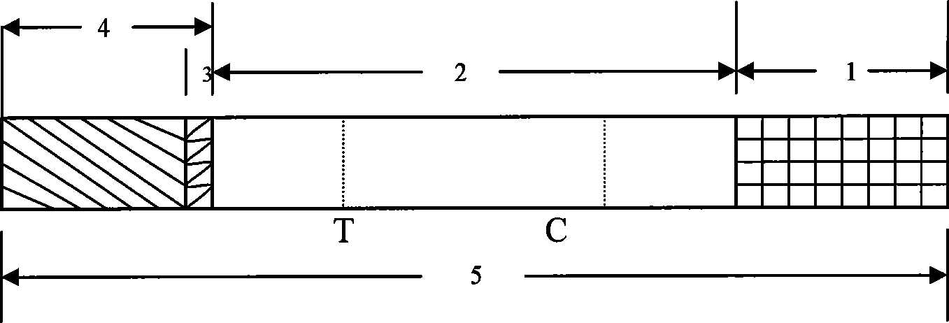
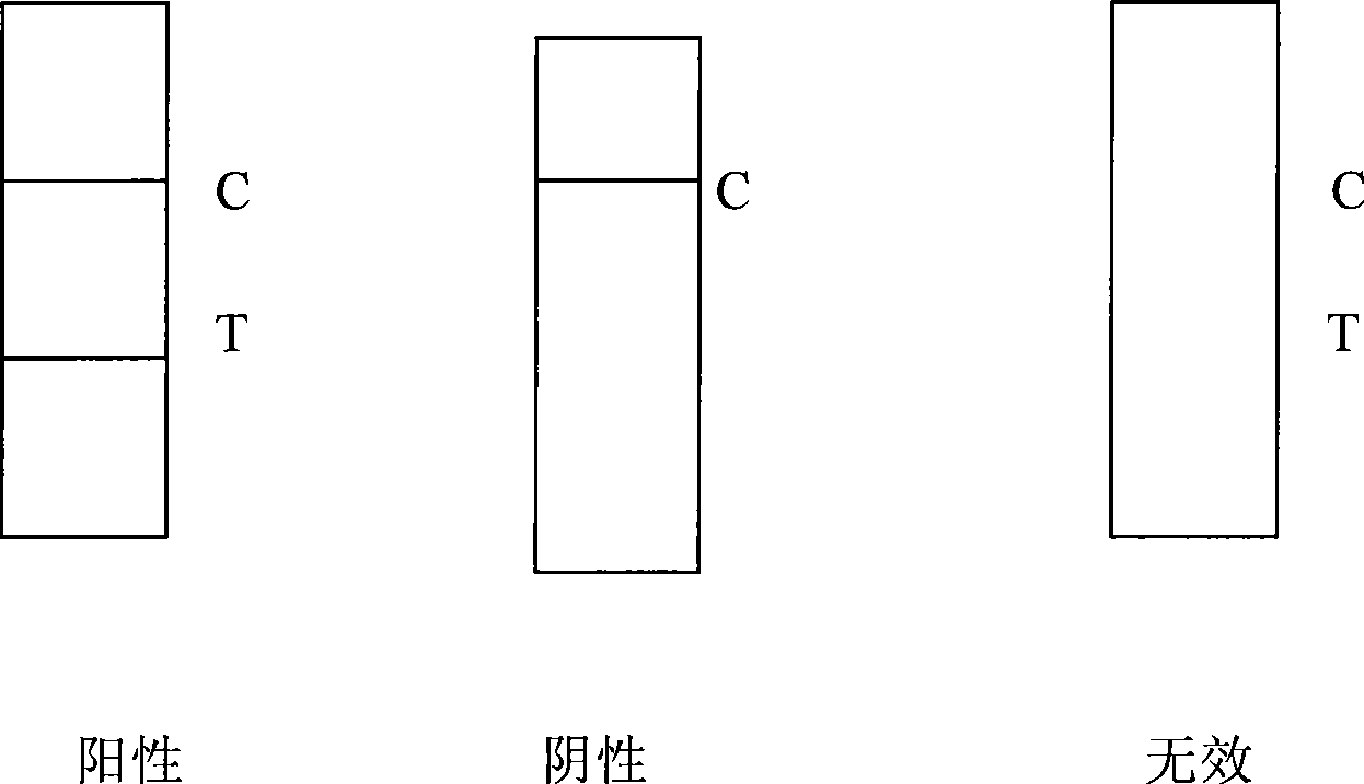
Test paper strip for detecting PRRSV antibody colloidal gold, method for making same and applications
A technology for detecting porcine PRRS virus and test strips, which is applied to measuring devices, instruments, scientific instruments, etc., can solve the problems of long maintenance time of N protein antibodies, and achieve the effects of saving manpower and material resources, easy promotion, and clear and easy-to-distinguish results
- Summary
- Abstract
- Description
- Claims
- Application Information
AI Technical Summary
Problems solved by technology
Method used
Image
Examples
Embodiment 1
[0047] Cloning and expression of embodiment 1 porcine blue ear virus N protein
[0048] (1) Obtaining the target gene
[0049] The genome of porcine PRRS virus was provided by Beijing Institute of Animal Husbandry and Veterinary Medicine. The pET-32a(+) expression vector and PMD-18T were purchased from Novagen. According to the characteristics of the pET-32a (+) expression vector, design primers with restriction endocoenzymes BamH I and Xho I restriction sites at both ends:
[0050] Upstream is 5′C GGATCC ATG CCAAATAAC AAC G3′
[0051] Downstream is 5'G CTC GAG TCA TGC TGA GGG TGA T3'
[0052] The target fragment N was amplified, and the amplification conditions were: denaturation at 94°C for 3 minutes, 45s at 94°C, annealing at 56°C for 90s, extension at 72°C for 90s, 30 cycles, and finally extension at 72°C for 10 minutes.
[0053] (2) Cloning of the target gene and screening of positive recombinants
[0054] After electrophoresis, the PCR amplified product was recovere...
Embodiment 2
[0061] Embodiment 2: the development of the monoclonal antibody cell line of porcine blue ear disease virus N protein
[0062] (1) Myeloma cells
[0063] SP2 / 0 myeloma cells: resuscitate SP2 / 0 cells stored in a liquid nitrogen tank, and culture them in DMEM medium containing 10% calf serum for 48-72 hours. Uniform, neatly arranged, logarithmically split, ready for fusion.
[0064] (2) Immunized mice
[0065] After the prepared genetically engineered antigen was taken out from the -20°C low-temperature refrigerator and dissolved, it was injected subcutaneously into BALB / C mice at multiple points (0.2ml / mouse) at intervals of 10 days. A total of 3 times of immunization. Three days before the fusion, mice were challenged with 0.15 ml of antigen in the peritoneal cavity.
[0066] (3) cell fusion
[0067] Fusion agent PEG (molecular weight 1500, produced in Japan); culture medium: 10% calf serum DMEM. Lymphocytes of SP2 / 0 cells and immunized BALB / C mice were divided into 2×10...
Embodiment 3
[0073] Embodiment 3: Preparation and assay of the monoclonal antibody of porcine blue ear disease virus N protein
[0074] (1) Monoclonal antibody ascites preparation:
[0075] Mice: SPF grade mice, no rodent virus contamination after inspection, if the animals are found to be unhealthy, bitten or infected during the ascites production process, they should be discarded.
[0076] Expanded culture of cell lines: Take one tube of the production batch of cells to resuscitate, add nutrient solution to expand the culture, and one production cell is only used once, and will not be frozen.
[0077] Inoculation of hybridoma cell lines: preparation of ascites should be carried out under sterile conditions, before injection of hybridoma cells, each mouse was intraperitoneally injected with liquid paraffin 0.5ml. One week later, each mouse was intraperitoneally injected with 1-3×10 hybridoma cells. 6 / 0.2ml.
[0078] Ascites collection: 7 to 10 days after injecting the cell line, or th...
PUM
 Abstract
Description
Claims
Application Information
 - R&D
- Intellectual Property
- Life Sciences
- Materials
- Tech Scout
- Unparalleled Data Quality
- Higher Quality Content
- 60% Fewer Hallucinations
Browse by: Latest US Patents, China's latest patents, Technical Efficacy Thesaurus, Application Domain, Technology Topic, Popular Technical Reports.
© 2025 PatSnap. All rights reserved.Legal|Privacy policy|Modern Slavery Act Transparency Statement|Sitemap|About US| Contact US: help@patsnap.com


