Encephalic angioma image recognizing and detecting method based on framework characteristic

A cerebral hemangioma and detection method technology is applied in the field of identifying and detecting cerebral hemangioma images based on skeleton features and constructing a computer-aided diagnosis system for cerebral hemangioma, which can solve the problem of low utilization value of cerebral hemangioma features, high missed detection rate, Hemangioma features are difficult to extract and other problems, to achieve the effect of fast speed, low missed detection rate, and accurate results

Inactive Publication Date: 2009-05-06
SUZHOU UNIV
View PDF0 Cites 11 Cited by
  • Summary
  • Abstract
  • Description
  • Claims
  • Application Information

AI Technical Summary

Problems solved by technology

[0005] The present invention provides a method for recognizing and detecting cerebral hemangioma images based on skeleton features, which is used to solve the problem that the existing pattern recognition method is difficult

Method used


Image

  • Encephalic angioma image recognizing and detecting method based on framework characteristic
  • Encephalic angioma image recognizing and detecting method based on framework characteristic
  • Encephalic angioma image recognizing and detecting method based on framework characteristic

Examples


Embodiment 1

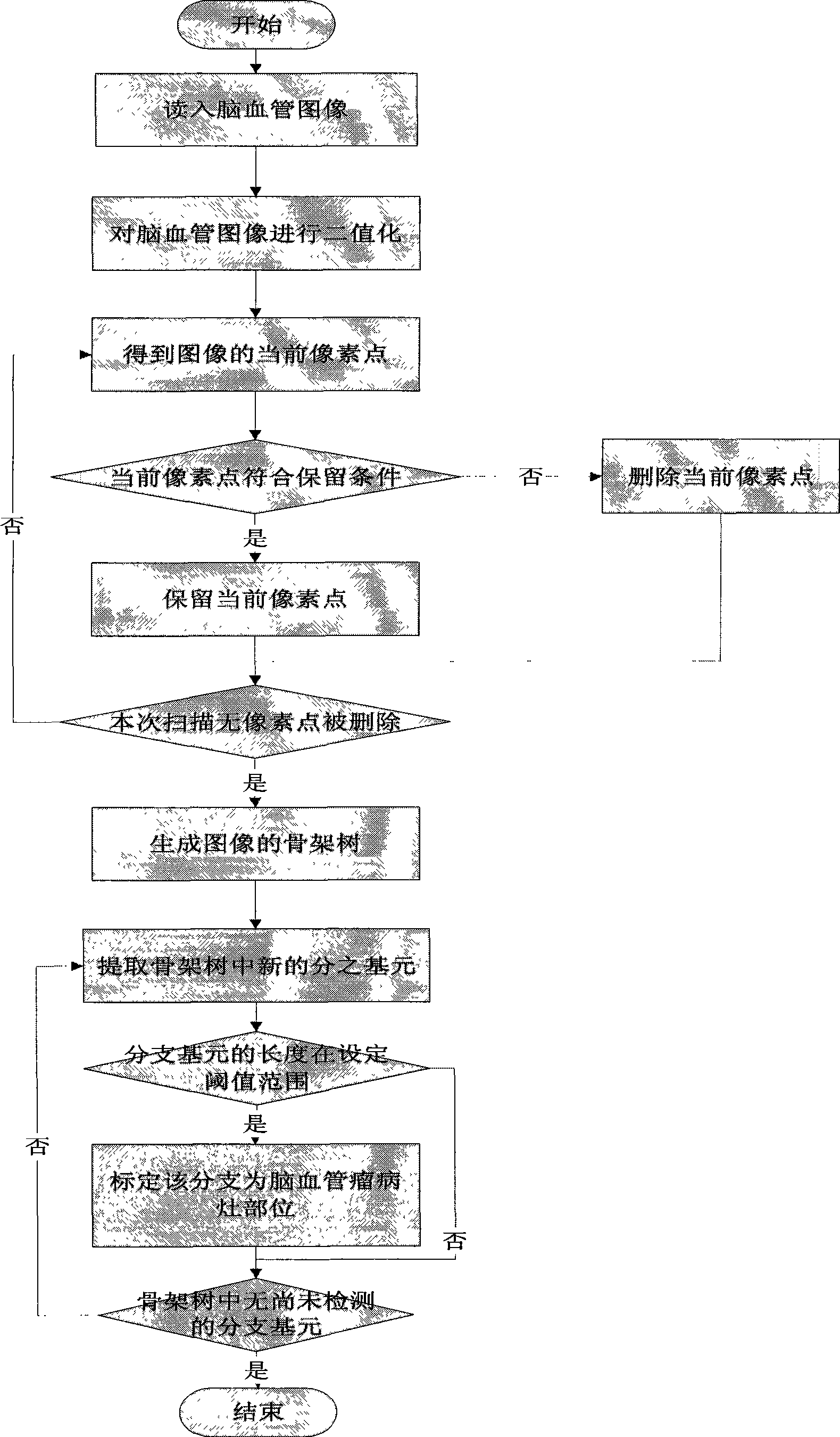
[0027] Embodiment one: figure 1 It is a flowchart of a method for detecting a cerebral hemangioma based on skeleton features, and the data file (picture file) is a picture of a cerebral blood vessel conforming to the BMP format.

[0028] (1) Binarization of the original image: the original image of the cerebrovascular is a DSA image of the cerebrovascular, and the format of the image conforms to the DICOM3.0 standard. Each DSA image is decomposed into DSA sequence images by using image processing software (in this embodiment, the DICOM software developed by the Institute of Intelligent Information Processing and Application of Soochow University) is used, and they are saved in BMP format. Binarize the grayscale image obtained in BMP format;

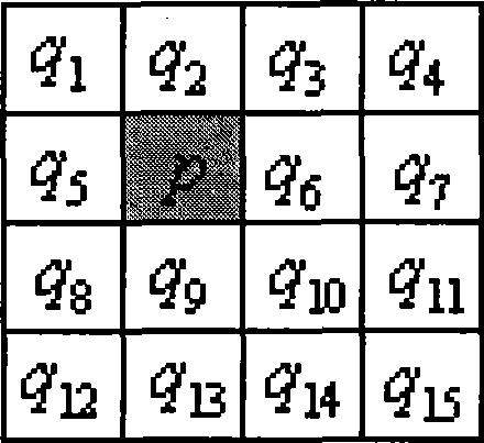
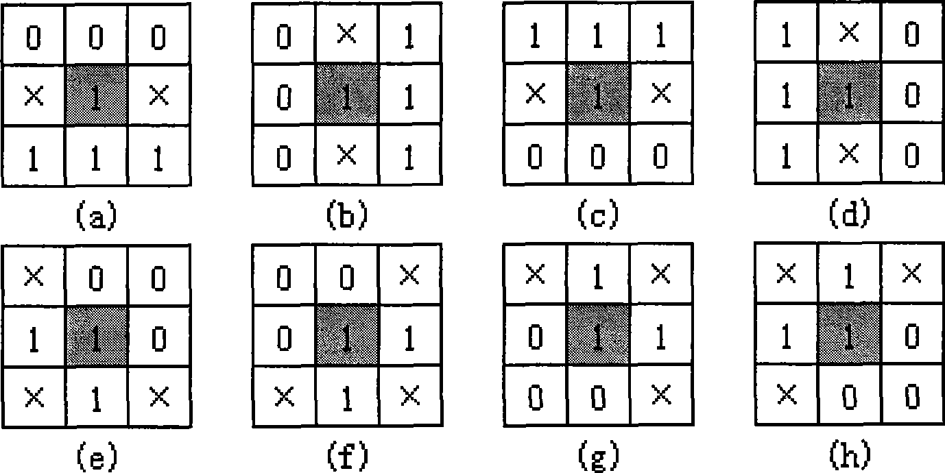
[0029] (2) Skeleton extraction: use an improved thinning algorithm to extract the skeleton from the binarized image obtained in step (1). This embodiment adopts an improved OPTA (one-pass thinning algorithm) thinning method. After thinn...


PUM


Abstract

The invention discloses a method for identifying and detecting encephalofacial angiomatosis image on a basis of skeleton characteristics. The method comprises the following steps: (1) performing binaryzation to a gray level image; (2) extracting a skeleton tree and obtaining a single-pixel curve image; (3) extracting a skeleton structure unit, wherein a key point unit is a branch point and an endpoint in the skeleton tree image; a branch unit is a skeleton section which connects two key point units but does not pass through a third key point, wherein at least one of the two key point units is a unit of an endpoint and is an external branch unit; and (4) determining thresholds T1 and T2, wherein the length of the external branch unit to be detected is S; and judging the encephalofacial angiomatosis image according to the relationship between S and T1 as well as T2, wherein T1 is an integer ranging between 6 to 10 and T2 is an integer ranging between 14 to 18. Due to the adoption of the method of the invention, time complexity of arithmetic is low, the result is precise, and good assistance can be provided for a doctor to diagnose the encephalofacial angiomatosis.

Description

technical field [0001] The invention belongs to the field of pattern recognition in medical image processing technology, relates to a method for recognizing and detecting cerebrovascular tumor images, in particular to a method for recognizing and detecting cerebrovascular tumor images based on skeleton features, which can be used to build a cerebrovascular tumor computer Auxiliary diagnostic system. Background technique [0002] Cerebrovascular diseases, especially cerebrovascular tumors, are one of the leading causes of death and disability in the world, seriously threatening human health and life. With the continuous maturity and development of computer technology, the computer-aided diagnosis (CAD) system produced by the combination of information technology and medical imaging technology is playing an increasingly important role in the detection and treatment of cerebrovascular diseases. , has also become one of the research hotspots in medical imaging. In the computer...

Claims


Application Information

Patent Timeline
IPC IPC(8): G06K9/00G06K9/46A61B19/00A61B90/00
Inventor 崔志明吴健翟海涛孙晓平张广铭
Owner SUZHOU UNIV
Why Eureka
Try Eureka