Endoscope for whole-course visible abortion operation
A surgical and endoscope technology is applied in the field of endoscopes for gynecological abortion surgery and endoscopes for abortion surgery, which can solve the problems of unclear surgical field of view, affecting the surgical effect and safety, etc., so as to improve safety and improve Operation efficiency and success rate, simple structure effect
- Summary
- Abstract
- Description
- Claims
- Application Information
AI Technical Summary
Problems solved by technology
Method used
Image

Examples
Embodiment 1
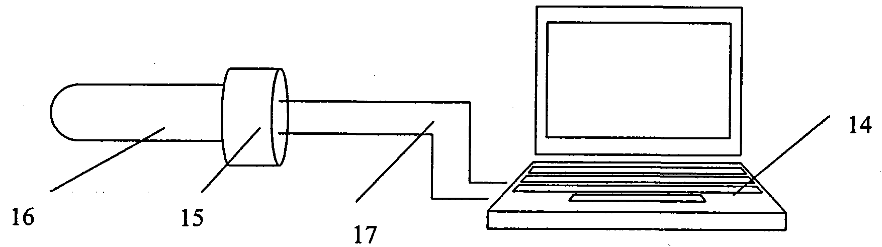
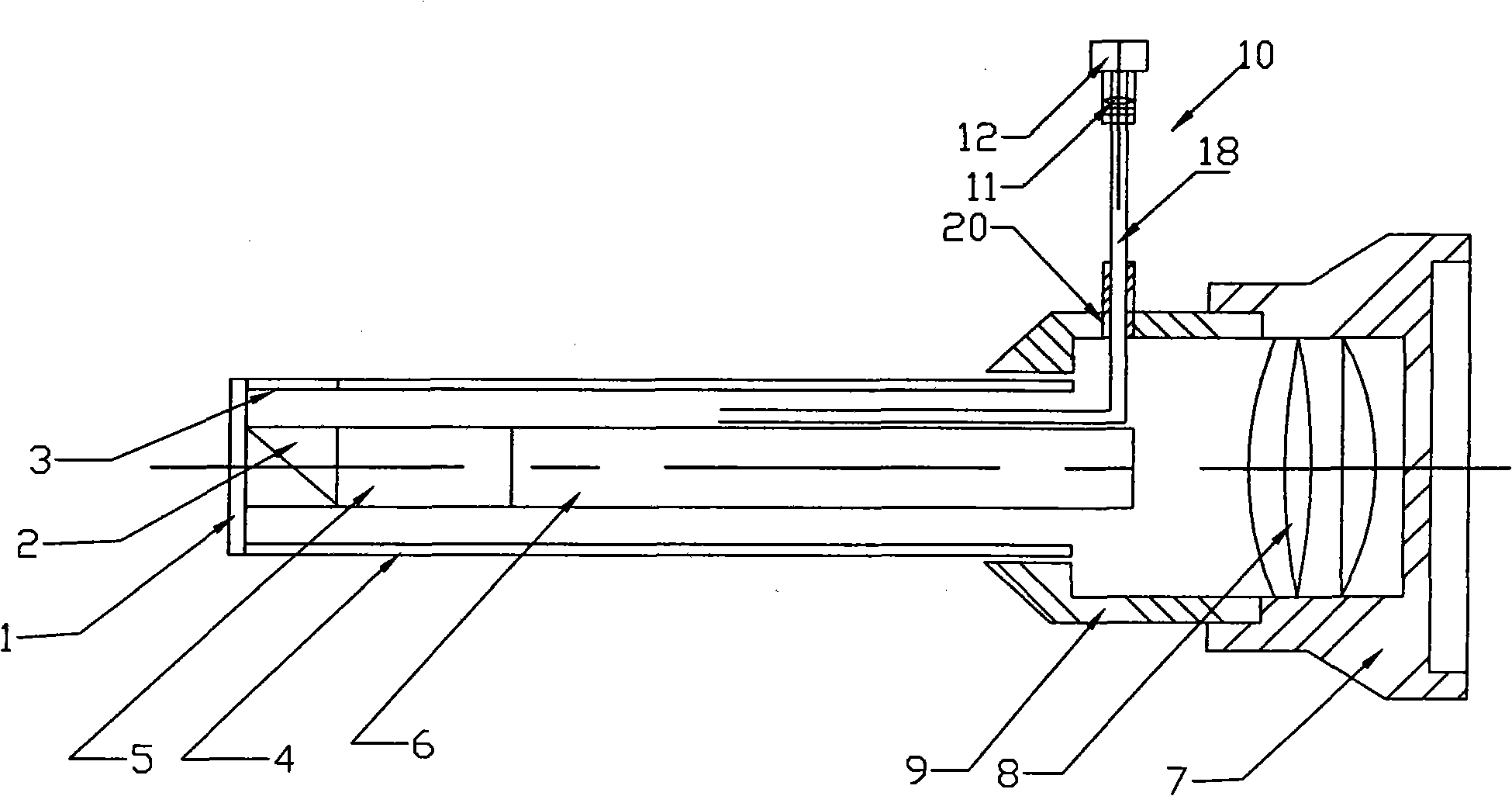
[0024] Such as figure 1 , 2 shown.
[0025] A full-range visible endoscope for abortion operation includes a computer 14, a CCD camera 15 and an endoscope assembly 16 capable of performing infrared imaging, such as figure 1 As shown, the photosensitive device of the CCD camera 15 is opposite to the eyepiece 8 of the endoscope assembly 16, the signal output end of the CCD camera 15 is connected with the signal input end of the computer 14 by the data line 17, and the light source of the endoscope assembly 16 is formed by the illumination light source (Its direction and structure are the same as those of the prior art) and an infrared imaging light source 10 capable of producing a wavelength greater than 700 nanometers (if the generated infrared wave energy is more than 1064 nanometers, the effect is better), the infrared imaging light source 10 is composed of a general lighting source 12 (the same as the existing endoscope illumination light source) plus an imaging light sour...
Embodiment 2
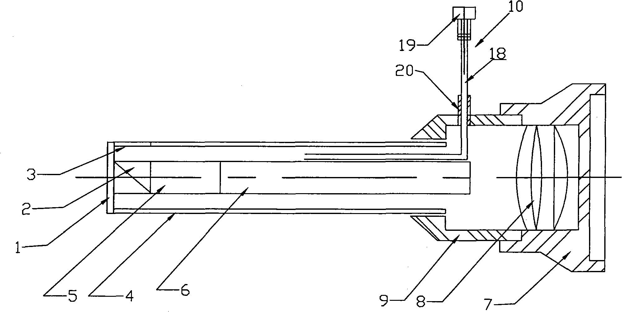
[0028] Such as figure 1 , 3 shown.
[0029] The difference between the present embodiment and the second embodiment is that the infrared imaging light source 10 directly adopts an infrared light spectrometer 19 capable of producing more than 700 nanometers (not less than 1064 nanometers is the best), and the lighting source before the operation needs to be configured separately. The same optical fiber 18 can be used with the infrared light spectrometer 19 to carry out light transmission (it can be properly converted during use), such as image 3 As shown, one illumination light path can also be provided separately, and the color filter 11 is omitted compared with the first embodiment, but the cost will increase. The specific implementation can also make the infrared light spectrometer work in the ordinary lighting mode first, and then switch to the state capable of emitting infrared light when bleeding occurs during the operation.
Embodiment 3
[0031] The difference between the present embodiment and the second embodiment is that the light spectrometer 19 is directly used as the light source for image capture, and the general illumination light source is omitted, which makes the whole structure simpler.
PUM
| Property | Measurement | Unit |
|---|---|---|
| Wavelength | aaaaa | aaaaa |
Abstract
Description
Claims
Application Information
 - R&D
- Intellectual Property
- Life Sciences
- Materials
- Tech Scout
- Unparalleled Data Quality
- Higher Quality Content
- 60% Fewer Hallucinations
Browse by: Latest US Patents, China's latest patents, Technical Efficacy Thesaurus, Application Domain, Technology Topic, Popular Technical Reports.
© 2025 PatSnap. All rights reserved.Legal|Privacy policy|Modern Slavery Act Transparency Statement|Sitemap|About US| Contact US: help@patsnap.com