Early-stage breast cancer nondestructive screening and imaging system

An imaging system and breast cancer technology, applied in the field of biomedical measurement and medical equipment, can solve the problems of inability to miniaturize equipment, expensive use and maintenance costs, high equipment cost, etc., to achieve miniaturization, simple machining, and easy operation easy effect

Inactive Publication Date: 2010-09-01
JIANGXI SCI & TECH NORMAL UNIV
View PDF3 Cites 17 Cited by
  • Summary
  • Abstract
  • Description
  • Claims
  • Application Information

AI Technical Summary

Problems solved by technology

[0003] At present, breast cancer screening mainly uses manual hand examination to assist other medical clinical detection methods. Commonly used ultrasound imaging uses reflection echo imaging when ultrasound waves encounter an interface with changing acoustic impedance during propagation in the body, but the imaging contrast of early-stage lesions is very low. Low; X-CT technology is an image formed by the different shadows produced by different parts of the tissue’s absorption of X-rays, but imaging harmful rays as information carriers may lead to an increase in the probability of cancer; MRI is used Coil detection technology obtains tissue relaxation information and proton density information imaging technology, which has no damage to the human body and can realize functional imaging, but the equipment cost is high, and the use and maintenance costs are expensive, causing a large economic burden to patients, and the equipment cannot miniaturization
At present, there is no low-cost, scalable, and especially suitable for early breast cancer detection and imaging in the clinical field of medicine

Method used


Image

  • Early-stage breast cancer nondestructive screening and imaging system
  • Early-stage breast cancer nondestructive screening and imaging system
  • Early-stage breast cancer nondestructive screening and imaging system

Examples


Embodiment 1

[0037] Example 1 An imaging system for nondestructive screening of unilateral early breast cancer

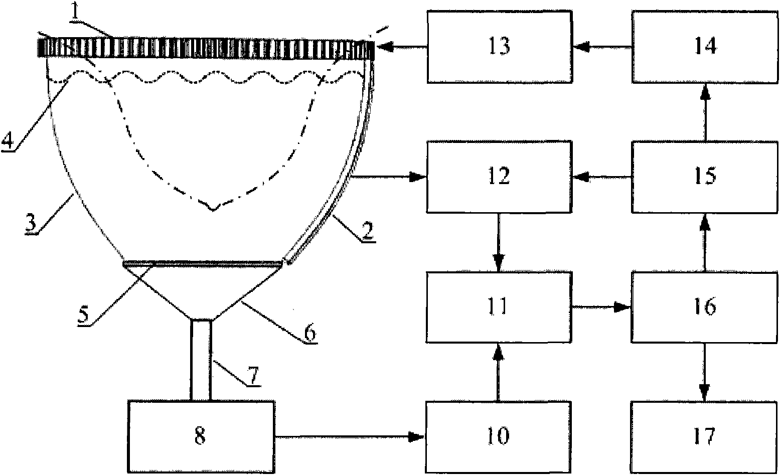
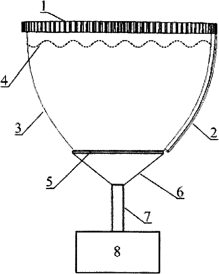
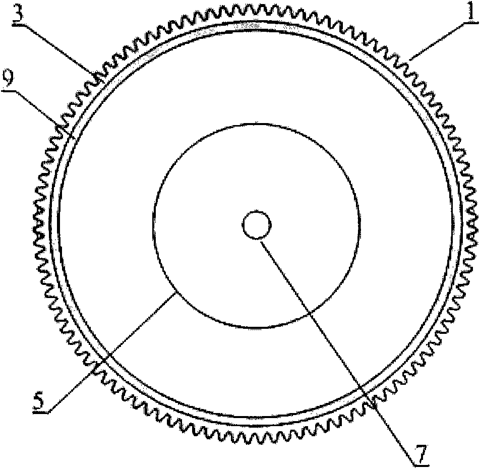
[0038] The structure of this embodiment is as Figure 1-4 As shown, the names of the components are: 1. Circular gear, 2. Arc-shaped ultrasonic array, 3. Bowl-shaped arc-shaped housing, 4. Ultrasonic coupling liquid, 5. Protective film, 6. Horn antenna, 7. Waveguide , 8. Microwave generator, 9. Elastic sealing ring, 10. Frequency divider, 11. Data acquisition circuit, 12. Preprocessing circuit, 13. Stepping motor, 14. Driver, 15. Digital I / O card, 16 .Computer, 17. Monitor.

[0039] Wherein the microwave generator 10 selects the BW-1200HPT of the No. 206 Research Institute of China Ordnance Industry for use, which can emit pulsed microwaves with a frequency of 1.2 GHz, and the pulse width is optional at 0.5 or 1 us; the data acquisition circuit 11 is an 8-channel synchronous sampling channel High-speed digitizer PCI-5105 (NI, the United States); curved ultrasonic array 2 is th...

Embodiment 2

[0051] An imaging system for non-destructive screening of bilateral breast early breast cancer

[0052] The structure of this embodiment is similar to Embodiment 1, the difference is:

[0053] 1) It also includes a fixed unit mainly composed of a workbench 18 . There are two coaxial circular through-holes 19 on the workbench 18, directly below each of the circular through-holes 19 is a thermoacoustic excitation and sensing unit, each thermoacoustic excitation and sensing unit Circular gear 1 in all is rotatably connected with workbench 18.

[0054] 2) A stepper motor 13 is meshed with the circular gears 1 in the two thermoacoustic excitation and sensing units at the same time.

[0055] 3) A frequency divider 10 is simultaneously connected to the wires of the microwave generators 8 in the two thermoacoustic excitation and sensing units.

[0056] 4) In each thermoacoustic excitation and sensing unit, an arc-shaped ultrasonic array 2 whose curvature matches the bowl-shaped arc...

Embodiment 3

[0059] An imaging system for non-destructive screening of bilateral breast early breast cancer

[0060] The structure of this embodiment is similar to Embodiment 2, the difference is:

[0061] The circular gear 1 in each thermoacoustic excitation and sensing unit is engaged with a stepping motor 13 respectively. Two stepping motors are connected with driver 14 wires simultaneously. The side wall of each bowl-shaped arc-shaped housing 3 is inlaid and fixed with two arc-shaped ultrasonic arrays 2 with an included angle of 90 degrees. Each arc-shaped ultrasonic array 2 contains 256 array elements, and the slits between the array elements are as wide as possible. It is 0.03mm, its center frequency is 2.5MHz, the relative bandwidth is 75%, and the area is 80mm×10mm×0.8mm.

[0062] The operation steps of the imaging system are the same as those described in Embodiment 1. The two bowl-shaped arc-shaped shells can rotate independently, and more scans can be performed on breasts with...


PUM


Abstract

The invention discloses an early-stage breast cancer nondestructive screening and imaging system, which mainly comprises circular gears, an annular ultrasonic array, a bowl-shaped annular shell, ultrasonic coupling liquid, a protective film, a horn antenna, a waveguide tube, a microwave generator, a frequency divider, a data acquisition circuit, a preprocessing circuit, a stepping motor, a driver, a digital I/O card, a computer and a display. The working process of the system is that: detected mammary glands are radiated by pulse microwaves to generate thermo-acoustic signals which are received by the annular ultrasonic array and are finally acquired by the computer; the driver drives the annular ultrasonic array to rotate to a next position around detected biological tissues; and the steps of acquisition and rotation are repeated until the thermo-acoustic signals of enough positions are received, and the computer re-establishes three-dimensional thermo-acoustic images of the detectedtissues. The early-stage breast cancer nondestructive screening and imaging system can quickly and nondestructively realize the three-dimensional thermo-acoustic imaging of the detected mammary glands to provide one of important bases for the diagnosis of early-stage breast cancer.

Description

technical field [0001] The invention relates to the technical fields of biomedical measurement and medical equipment, in particular to an early breast cancer nondestructive screening imaging system. Background technique [0002] Breast cancer is the most common malignant tumor that seriously affects women's physical and mental health and even threatens their lives. According to statistics, the incidence rate accounts for 7-10% of all kinds of malignant tumors in the whole body. About 1.2 million women are diagnosed with breast cancer every year in the world , and 400,000 women died of it, and the number is increasing at a rate of 2% to 3% every year. It is estimated that the annual number of new cases of breast cancer in the world will reach 1.4 million in 2010. Now in our country, breast cancer has accounted for the first place in the incidence of female tumors in some cities, such as Shanghai and Wuhan, Beijing, Tianjin, Harbin and other cities have also taken the second p...

Claims


Application Information

Patent Timeline
Patent Type & Authority Applications(China)
IPC IPC(8): A61B8/00
Inventor 曾吕明杨迪武刘国栋徐景坤
Owner JIANGXI SCI & TECH NORMAL UNIV
Why Eureka
Try Eureka