Method for preparing live animal eye model by retinal vein artificial blood vessel bypass operation

A retinal vein and artificial blood vessel technology, applied in medical science, veterinary instruments, veterinary surgery, etc., can solve problems such as limiting the research and development of intraocular retinal blood vessel ultra-microsurgery

Inactive Publication Date: 2010-10-06
WENZHOU MEDICAL UNIV
View PDF0 Cites 4 Cited by
  • Summary
  • Abstract
  • Description
  • Claims
  • Application Information

AI Technical Summary

Problems solved by technology

Due to the slender vascular diameter of the retina (retinal artery diameter is less than 0.1mm, retinal vein diameter is less than 0.2mm), the material of artificial blood vessels and t

Method used


Image

  • Method for preparing live animal eye model by retinal vein artificial blood vessel bypass operation
  • Method for preparing live animal eye model by retinal vein artificial blood vessel bypass operation
  • Method for preparing live animal eye model by retinal vein artificial blood vessel bypass operation

Examples


Embodiment Construction

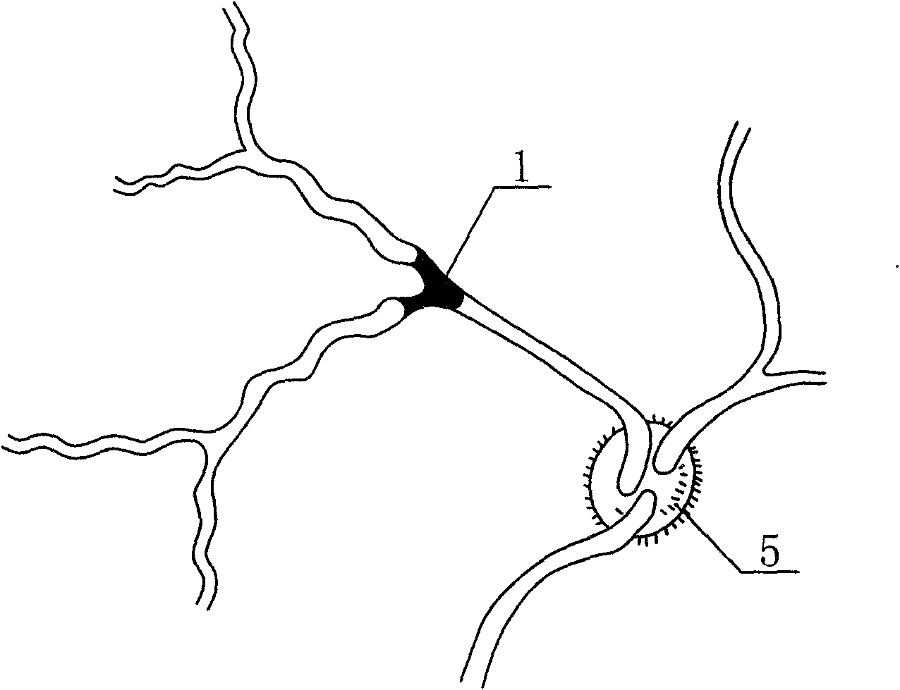
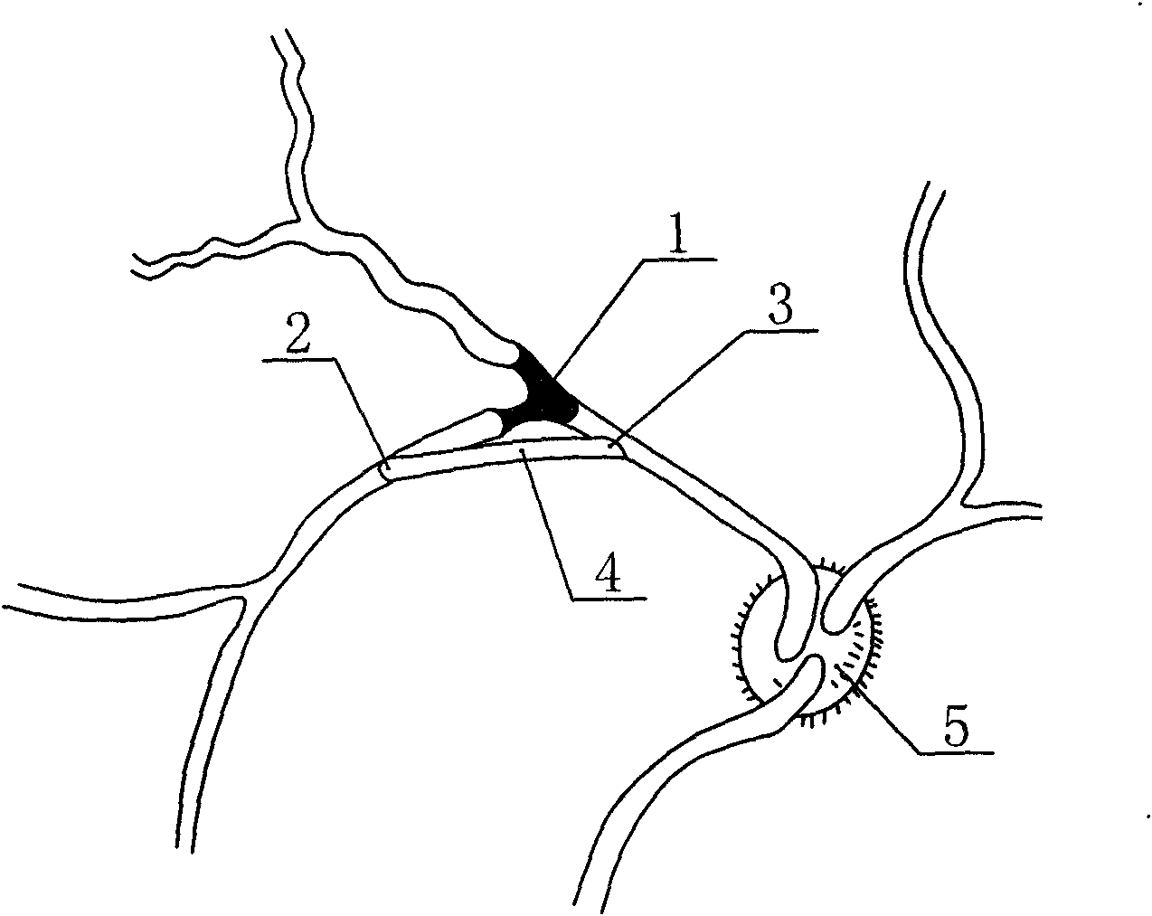
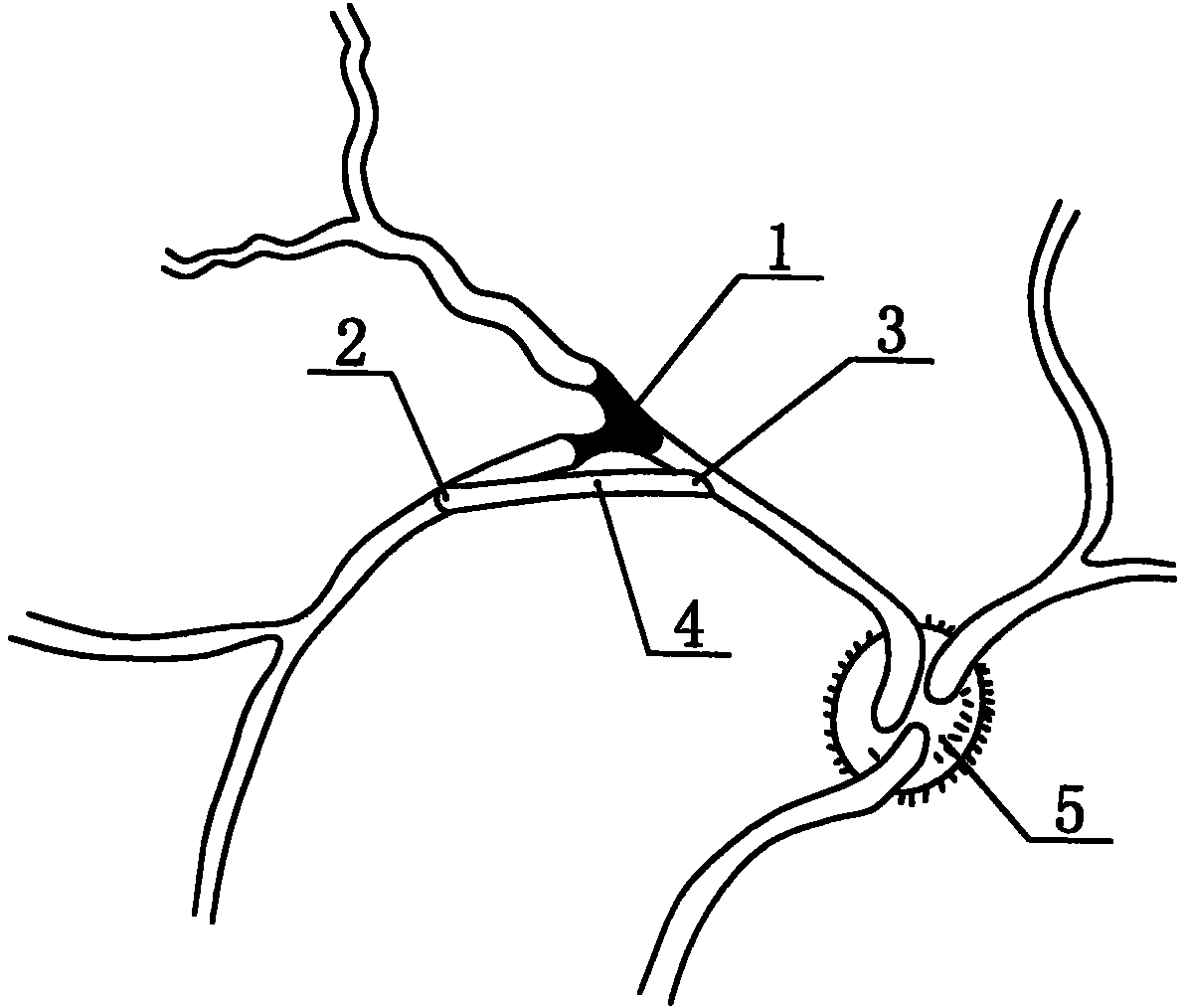
[0018] The experimental animals of the living animal eye model of the retinal vein artificial vascular bypass in the present invention can be selected from mammals such as pigs, dogs, sheep, monkeys, and the specific details of the living animal eye model of the present invention are introduced as an example below with miniature pigs for experiments. Production Method:

[0019] First, 2-month-old experimental minipigs were selected, and the anticoagulant drugs Aspirin enteric-coated sustained-release tablets 300 mg / day and warfarin sodium tablets 500 mg / day were given for five consecutive days before the operation. 5-7 days before the operation, puncture the anterior chamber with a 1ml syringe to extract 0.1ml of aqueous humor, and inject 0.5U / 0.05ml of plasmin and 50U / 0.05ml of plasmin into the vitreous cavity through a 30G needle at 2.5mm behind the corneal limbus in the superior temporal quadrant. ml hyaluronidase can induce complete posterior vitreous detachment, which fac...


PUM


Abstract

The invention relates to a method for preparing a live animal eye model by a retinal vein artificial blood vessel bypass operation. The method comprises the following steps of: selecting experimental pigs, dogs, sheep and monkeys, artificially causing branch retinal vein occlusion in advance and then performing vitrectomy; setting a point on each of blood vessels on two sides of the occlusion as a starting point and an end point of a vascular bypass respectively, designing a bending curvature, length and acute end angles by taking a macromolecular material microtubule as an artificial blood vessel; and after the artificial blood vessel is introduced into an eye, puncturing vessel walls at the position of the set starting point and the set end point by two acute ends and inserting the two acute ends into the artificial blood vessel sequentially, wherein punctured pores are automatically closed or are promoted to be closed by performing electric coagulation on the blood vessel if necessary. By establishing the live animal eye model used for the research on the time, the effect and the security of curing the branch retinal vein occlusion by the retinal vein artificial blood vessel bypass operation, the method lays a research foundation for developing a new clinical surgery method.

Description

technical field [0001] The invention relates to a method for making a living animal eye model used in the research of surgical treatment of retinal vein occlusive disease in experimental medicine, in particular to a method for making a living animal eye model for retinal vein artificial blood vessel bypass. Background technique [0002] Branch Retinal Vein Occlusion (BRVO) is the second common retinal vascular disease that causes visual impairment after diabetic retinopathy. Severe cases can lead to blindness. It has always been the focus of research in the field of fundus diseases. It is generally believed that the disease is caused by retinal vein reflux caused by retinal vein thrombosis, but the specific cause and pathological mechanism are not completely clear. At present, the commonly used retinal laser photocoagulation and vitrectomy surgery only focus on the ischemic and hypoxic complications after branch vein occlusion, not the treatment of the cause. However, some ...

Claims


Application Information

Patent Timeline
IPC IPC(8): A61D1/00
Inventor 沈丽君陈亦棋吴炜
Owner WENZHOU MEDICAL UNIV
Why Eureka
Try Eureka