Method and device for assisting in gene transfer by utilizing electroporation

A technology of gene transfer and electroporation, which is applied in the field of ophthalmology, can solve the problems of complex cell hierarchy structure and difficult design and application, and achieve the effect of convenient operation, avoiding cell immunogenicity and potential pathogenicity, and improving stability

Active Publication Date: 2012-11-07
EYE & ENT HOSPITAL SHANGHAI MEDICAL SCHOOL FUDAN UNIV
View PDF0 Cites 0 Cited by
  • Summary
  • Abstract
  • Description
  • Claims
  • Application Information

AI Technical Summary

Problems solved by technology

In contrast, its design and application in the eye is more difficult. First, the shape of the eyeball itself means that a simple flat electrode will not be able to do it; second, the cell hierarchy in the retinal tissue is complex, so the electroporation of the eye Dyeing requires special electrodes

Method used


Image

  • Method and device for assisting in gene transfer by utilizing electroporation
  • Method and device for assisting in gene transfer by utilizing electroporation
  • Method and device for assisting in gene transfer by utilizing electroporation

Examples


Embodiment 1

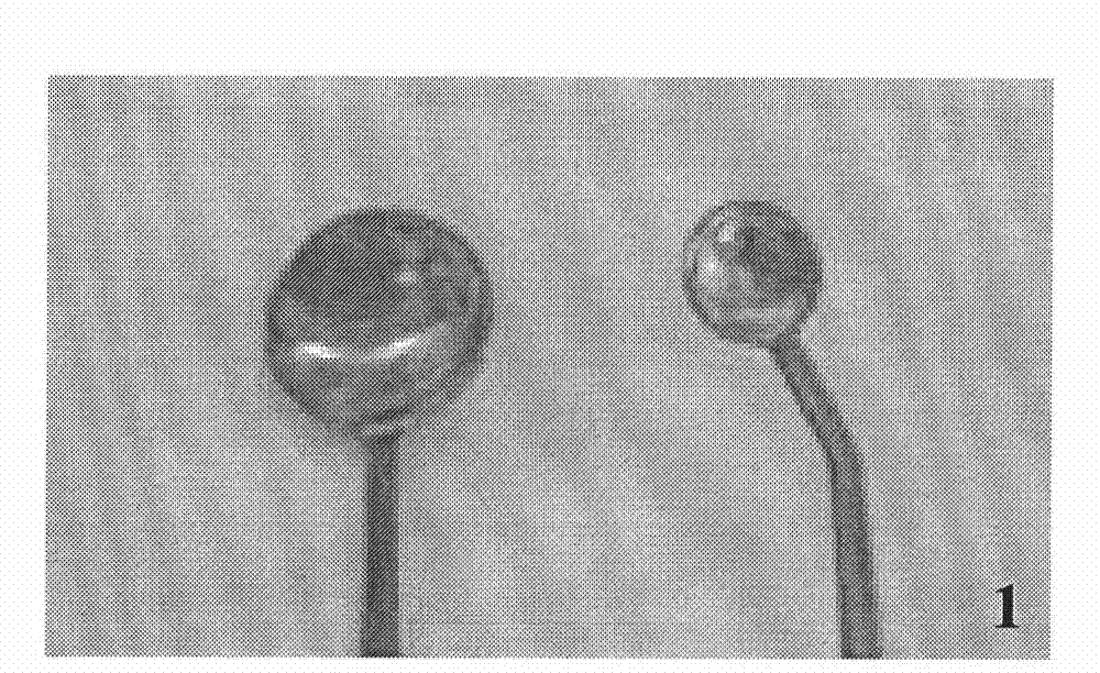
[0046] Example 1 Preparation of double spoon type special electrode device

[0047] On the basis of the conventional ophthalmic electrode device, gold is used as the conductive contact surface material of the electrode, copper-aluminum alloy is used as the support material, and the outer layer is coated with resin as the insulating layer, and the specifications of the obtained electrode shape are guaranteed by measurement, double spoon type electrode( figure 1 ) Its two poles are connected to the electrical stimulation generator through a plug, and the positive and negative poles can be adjusted as required. The cornea-side electrode of the double-spoon electrode is in the shape of a large round spoon, which is mainly in line with the area and curvature of the cornea; the electrode on the scleral side is slightly smaller, in the shape of a small round spoon, and is only placed at the plasmid transfer site, which can reduce the separation range of the retrobulbar area and Red...

Embodiment 2

[0049] 1. Construction of plasmid vectors

[0050] The existing plasmid pRc / CMV BDNF contains mouse BDNF cDNA, and the fragment length is 750bp. The primers of sequence 1 and sequence 2 (5'-ATG ACC ATC CTT TTC CTT ACT ATG-3' and 5'-CCA CTA TCT TCCCCT TTT AAT GG-3') were designed and synthesized. PCR reaction (94°C for 30s, 57°C for 30s, 72°C for 40s, 30 cycles) amplified the target gene, electrophoresed and recovered to obtain BDNF cDNA. Then, according to the instructions provided by NT-GFP Fusion TOPO TA Expression Kits, the connection of BDNF cDNA and pcDNA3.1 / NT-GFP-TOPO vector plasmid, transformation, plasmid extraction, and sequencing were carried out. After sequencing confirmation, use the Endofree Plasmid Purification Maxi kit to extract the plasmids and detect the OD260 / OD280 value of the obtained plasmids. If the ratio is greater than 1.8, it is qualified, and the qualified plasmids are diluted to a concentration of 2.5 μg / μl.

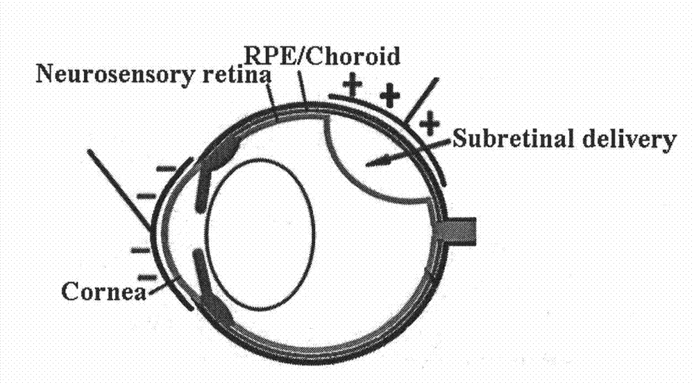
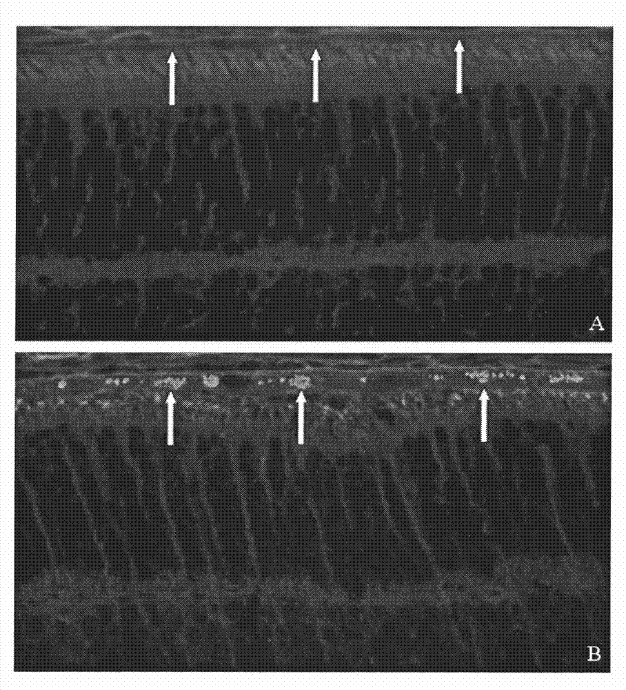
[0051] 2. Microsurgery experiments a...


PUM

PropertyMeasurementUnit
diameteraaaaaaaaaa

Abstract

The invention belongs to the technical field of ophthalmology, and relates to a method and a device for assisting in gene transfer by utilizing electroporation. In the method and the device, a double-spoon electrode device is adopted, proper electric stimulation pulse conditions are determined, and green fluorescence protein-containing plasmids are transfected by a retinal pigment epithelial celllayer. The method of the invention has the advantages of directionally controlling the intraocular transfection of genes by using an electric field, simultaneously avoiding the shortcomings of cellular immunogenicity, potential pathogenicity, tumorigenicity and the like caused by viral gene transfer, reducing side injuries to eyeballs by adopting the double-spoon electrode device, simultaneously realizing the transfection of a plurality of plasmids, and compensating for the shortcomings of conventional methods, along with convenient operation, high efficiency, capability of being used as a reference for the gene transfer in ophthalmic researches, and the like.

Description

technical field [0001] The invention belongs to the technical field of ophthalmology and relates to a method for retinal gene transfer, in particular to a method for using electroporation to assist gene transfer into retinal pigment epithelial cell layer in vivo. Background technique [0002] Studies have shown that retinitis pigmentosa, proliferative vitreoretinopathy, and age-related macular degeneration, which mainly involve the retinal pigment epithelial cell layer, are one of the major blinding eye diseases in the world. Although their pathogenesis is different, including genetic defects, abnormal proliferation of retinal pigment epithelial cells and metabolic disorders, etc., the lesion sites involved are all located in the retinal pigment epithelial cell layer. [0003] Since the retinal pigment epithelial cell layer exists in the innermost layer of the retina in the closed posterior hemisphere of the eye, and the retina also has a blood-ocular barrier that blocks the...

Claims


Application Information

Patent Timeline
Patent Type & Authority Patents(China)
IPC IPC(8): C12N15/85
Inventor 莫晓芬张萌吴继红徐格致倪颖勤董京艳
Owner EYE & ENT HOSPITAL SHANGHAI MEDICAL SCHOOL FUDAN UNIV
Why Eureka
Try Eureka