Diagnosis and treatment integrated confocal hysteroscope system

A hysteroscopy and confocal technology, used in laparoscopy, diagnosis, endoscopy, etc., can solve the problems of increasing patient pain, wasting operation time, delaying patient treatment time, etc., saving operation time, improving accuracy and safety effect

Inactive Publication Date: 2011-04-20
GUANGZHOU BAODAN MEDICAL INSTR TECH
View PDF4 Cites 4 Cited by
  • Summary
  • Abstract
  • Description
  • Claims
  • Application Information

AI Technical Summary

Problems solved by technology

When the traditional rigid hysteroscope enters the patient's uterine cavity, the optical system can observe the real situation in the uterine cavity in real time. With the cooperation of the corresponding surgical instruments through the channel, the doctor can solve most of the patient's problems through the rigid hysteroscope. For gynecological lesions, for suspicious lesions such as tumors and inflammations in the uterine cavity or mucosa, doctors often need to access surgical instruments such as biopsy forceps

Method used


Image

  • Diagnosis and treatment integrated confocal hysteroscope system
  • Diagnosis and treatment integrated confocal hysteroscope system
  • Diagnosis and treatment integrated confocal hysteroscope system

Examples


Embodiment 1

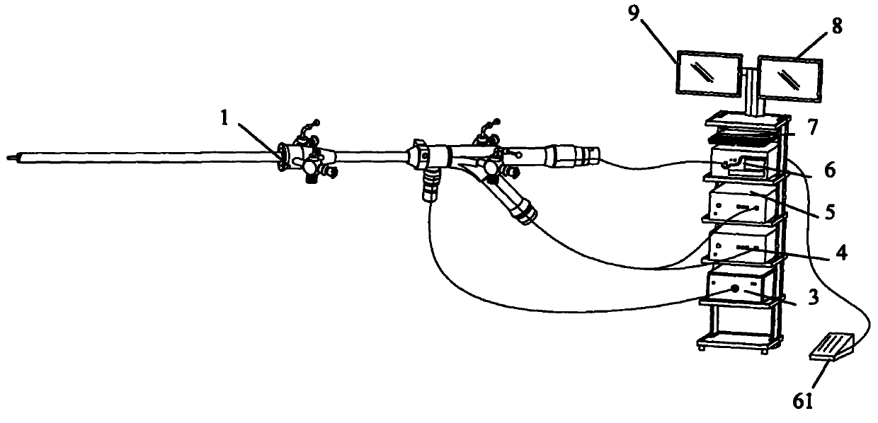
[0028] Such as figure 1 As shown, the diagnosis and treatment integrated confocal hysteroscope system of the present invention includes a rigid hysteroscope 1, a cold light source host 3 connected to the rigid hysteroscope, a camera host 4, and a confocal laser scanning microscope system host 5. Laser knife system host 6, keyboard 7 and monitors 8 and 9.

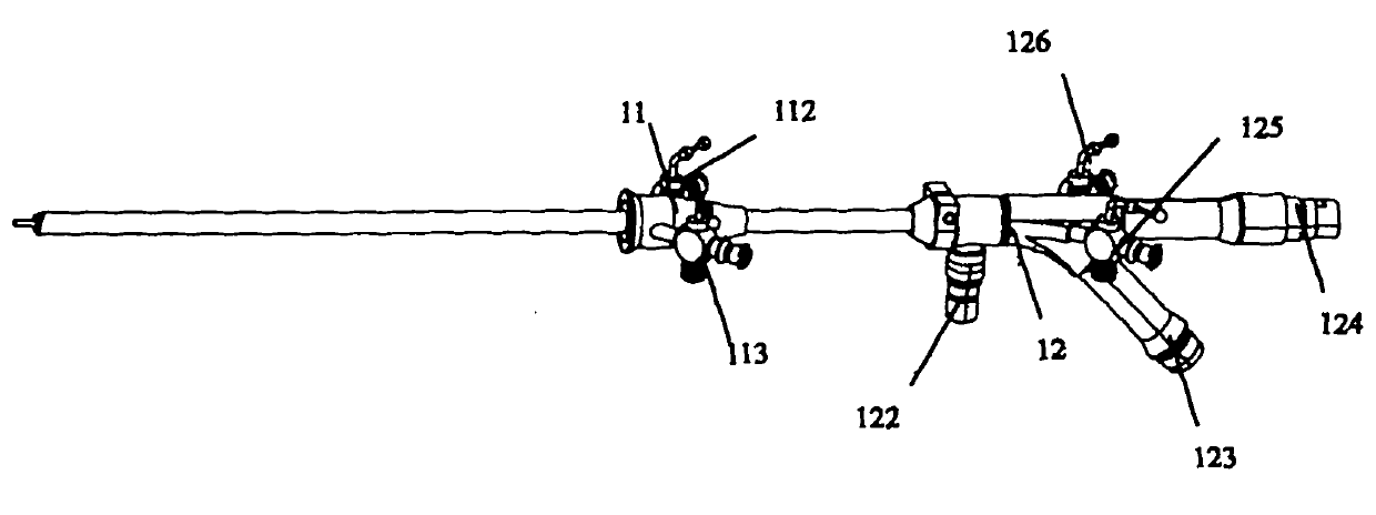
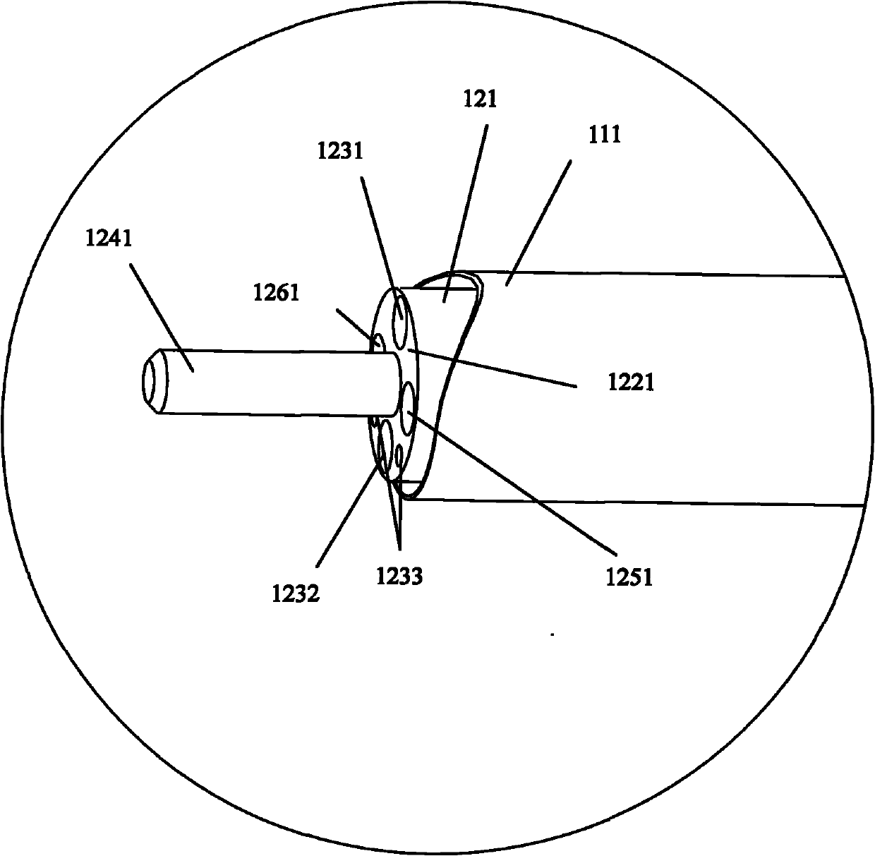
[0029] Such as figure 2 As shown, the rigid hysteroscope includes a main body endoscope part 12 and a hysteroscope sheath part 11 connected with the main body endoscope part 12 . The main body endoscope part 12 includes a rigid endoscope end part 121, a cold light source connector 122, an image data output port 123, a laser knife control interface 124, instrument channels 125 and 126; the hysteroscope sheath part 11 includes a sheath tube The main body, the water inlet channel 112 and the water outlet channel 113 communicated with the sheath tube main body, and the sheath tube end 111 arranged at the front end of the shea...

Embodiment 2

[0033] This embodiment is basically the same as the first embodiment above, the difference is:

[0034] Such as Figure 5 As shown, the diagnosis and treatment integrated confocal hysteroscope system of the present invention includes a rigid hysteroscope 2, a cold light source host 3 connected to the rigid hysteroscope, a camera host 4, and a confocal laser scanning microscope system host 5. Microwave knife system host 10, keyboard 7 and monitors 8,9.

[0035] Image 6 As shown, the rigid hysteroscope 2 includes a main body endoscope part 22 and a hysteroscope sheath part 21 connected with the main body endoscope part 22 . The main endoscope part 22 includes a rigid endoscope end 221, a cold light source connector 222, an image data output port 223, a laser knife control interface 224, and several instrument channels 225 and 226; the hysteroscope sheath part 21 includes The sheath tube main body, the water inlet channel 212 communicated with the sheath tube main body, the w...


PUM

PropertyMeasurementUnit
Outer diameteraaaaaaaaaa
Diameteraaaaaaaaaa
Lengthaaaaaaaaaa

Abstract

The invention belongs to the field of medical apparatus, and particularly relates to a diagnosis and treatment integrated confocal hysteroscope system. A hard endoscope end part of a hard hysteroscope is provided with a treatment device; and a treatment system host, which is correspondingly used in conjunction with the treatment device, is connected to the hard hysteroscope. Particularly, the diagnosis and treatment integrated confocal hysteroscope system organically combines the hard hysteroscope, a confocal laser scanning microscope and a laser knife system or the hard hysteroscope, the confocal laser scanning microscope and a microwave knife system. Through the clinical application of the diagnosis and treatment integrated confocal hysteroscope system, the effect of performing diagnosis and treatment at the same time can be achieved, an optical system and a confocal laser scanning microscopic system can be used for observing the macroscopic condition and microscopic structures of a uterine cavity wall and pathologic changes in a uterine cavity respectively, and the laser knife system or the microwave knife system can perform laser treatment or microwave treatment on the pathologic changes under the direct vision of a monitor. By suing the diagnosis and treatment integrated confocal hysteroscope system to perform a hysteroscope operation, the problems in the diagnosis and the treatment of a patient can be solved, the frequent replacement of the endoscope can be avoided, a great deal of operation time can be saved, the paint of the patient can be alleviated, and the accuracy and the safety of the operation can be further improved.

Description

technical field [0001] The invention belongs to the field of medical devices, and in particular relates to a confocal hysteroscope system integrating diagnosis and treatment for hysteroscopic surgery. Background technique [0002] At present, hysteroscopy is widely used in gynecological diagnosis and treatment, and it is an important treatment tool for intrauterine tumors, polyps and other lesions. When the traditional rigid hysteroscope enters the patient's uterine cavity, the optical system can observe the real situation in the uterine cavity in real time. With the cooperation of the corresponding surgical instruments through the channel, the doctor can solve most of the patient's problems through the rigid hysteroscope. For gynecological lesions, for suspicious lesions such as tumors and inflammations in the uterine cavity or mucosa, doctors often need to access surgical instruments such as biopsy forceps to obtain tissues and send them for inspection. The process of send...

Claims


Application Information

Patent Timeline
IPC IPC(8): A61B1/313A61B18/18A61B18/22A61B1/04A61B1/07
Inventor 乔铁
Owner GUANGZHOU BAODAN MEDICAL INSTR TECH
Why Eureka
Try Eureka