Method for noninvasively and quantitatively measuring local blood flows of human organs

A technology for quantitative determination of human organs, applied in the field of biological and medical imaging, can solve problems such as difficult application, deviation of blood flow value distribution, distortion of input function combination, etc., to achieve the effect of high speed, high accuracy, and little influence of noise

Inactive Publication Date: 2011-05-25
NANJING UNIV OF INFORMATION SCI & TECH
View PDF3 Cites 9 Cited by
  • Summary
  • Abstract
  • Description
  • Claims
  • Application Information

AI Technical Summary

Problems solved by technology

However, these methods have such problems: a) a fast image acquisition scheme is required to reflect the time curve characteristics of the input function, especially the rapid changes in the early stage after FDG injection; b) the local volume effect needs to be effectively resolved; c) correction is required from the surrounding Spillover of brain regions of selected ROIs to these ROIs
However, since these specific reference a

Method used


Image

  • Method for noninvasively and quantitatively measuring local blood flows of human organs
  • Method for noninvasively and quantitatively measuring local blood flows of human organs
  • Method for noninvasively and quantitatively measuring local blood flows of human organs

Examples


Embodiment Construction

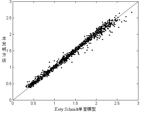
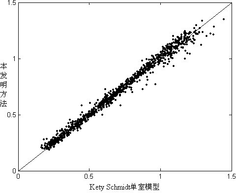
[0021] Below in conjunction with accompanying drawing introduces with simulated data (according to the experimental data parameter of existing literature report with the PET / H that computer randomly produces 2 15 (2 dynamic scanning data) the comparison of the present invention and internationally recognized gold standard Kety-Schmidt single-chamber model method determination result that carry out:

[0022] figure 1 , figure 2 The whole brain (absolute value of blood flow is taken as 0.5 ml / ml / min, this value is taken from the experimental results reported in the literature, and this value is the average value of all subjects in the existing experiments in the literature.) In the reference area, when the noise level is 1, the brain pixel by pixel is compared with the Kety-Schmidt single-chamber model method ( figure 1 ) and absolute ( figure 2 ) regression comparison of blood flow values. The scatter points in the figure correspond to the results of each pixel, and the ...


PUM


Abstract

The invention relates to a method for noninvasively and quantitatively measuring local blood flows of human organs. The method comprises the following steps of: performing positron emission tomography/fluorodeoxyglucose (PET/H2150) dynamic scanning on a human body, wherein scanned parts are determined according to circumstances; selecting the whole brain or other organs as reference areas as required, or selecting any parts of the brain or other organs as reference areas, and directly applying a formula; determining a relative local blood flow P1 of each part in the brain or other organs, wherein the part is not the same part as a contained area, and the P1 is automatically taken as 1 when the areas are completely overlapped; and performing normalization processing on the whole brain or the whole organs after the value of the relative local blood flow P1 of each part of the selected brain or other organs is solved. By the method, the per-pixels of the analyzed organs can be quantitatively determined, functional images of the local blood flow can be generated and input functions are not needed, so that blood drawing is not needed, and complicated correction is not needed. The method has high accuracy and facilitates practical application.

Description

technical field [0001] The present invention relates to PET / H of local blood flow in human brain and other organs 2 15 (0) Quantitative measurement method, which can perform quantitative measurement on the analyzed organ pixel by pixel, and then generate functional images of local blood flow, which belongs to the field of biological and medical imaging. Background technique [0002] Using Positron Radionuclides 15 O labeled water (i.e. H 2 15 O) Positron Emission Tomography (PET) method as imaging agent (abbreviated as PET / H 2 15 O method), is for intravenous H 2 15 After O, PET dynamic scanning is performed, and then the Kety-Schmidt single-chamber model and other methods are used for quantitative calculation of local blood flow pixel by pixel, so that images of local blood flow can be generated. [0003] In the early quantitative computing research, the input function needs to be measured by dynamically drawing arterial blood or arterialized venous blood. Although...

Claims


Application Information

Patent Timeline
IPC IPC(8): A61B6/00
CPCA61B6/507
Inventor 吴义根陈英茂
Owner NANJING UNIV OF INFORMATION SCI & TECH
Why Eureka
Try Eureka