Detector in nuclear medicine diagnosis device and using method thereof

A technology of a diagnostic device and a detector, applied in the field of nuclear medicine, can solve the problems of a large increase in the cost of a PET detector, and achieve the effect of improving the spatial resolution

Active Publication Date: 2011-07-06
北京锐视康科技发展有限公司
View PDF3 Cites 5 Cited by
  • Summary
  • Abstract
  • Description
  • Claims
  • Application Information

AI Technical Summary

Problems solved by technology

[0006] Embodiments of the present invention provide a detector in a nuclear medicine diagnostic device and a method for using the same, which is used

Method used


Image

  • Detector in nuclear medicine diagnosis device and using method thereof
  • Detector in nuclear medicine diagnosis device and using method thereof
  • Detector in nuclear medicine diagnosis device and using method thereof

Examples


Embodiment approach

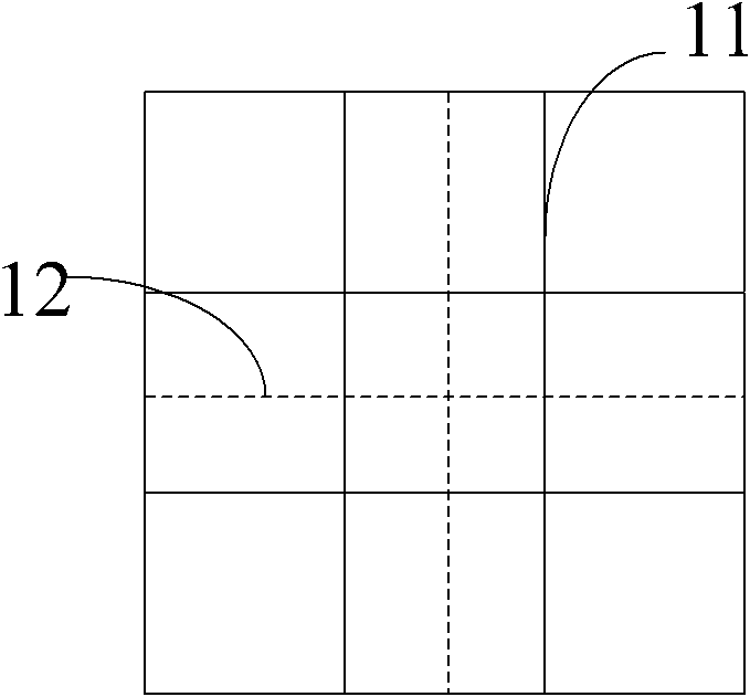
[0025] (1) As shown in Figure 3 (a), the area ratio of the coupling surface of the scintillator crystal 11 to the coupling surface of the photodetector 12 is less than 1:1, the coupling surface of the scintillator crystal 11 is a square, and the coupling surface of the scintillator crystal 11 is square. The coupling surface of the photodetector 12 is square, and the side length of the scintillator crystal is 2 / 3 of the side length of the detector. The photodetector 12 can be coupled with four scintillator crystals 11 , and the scintillator crystal 11 needs to be fully coupled through at least one photodetector 12 . Specifically, the scintillator crystal realizes complete coupling through 2 or 4 adjacent photodetectors.

[0026] (2) As shown in Figure 3 (b), the area ratio of the coupling surface of the scintillator crystal 11 to the coupling surface of the photodetector 12 is less than 1:1, the coupling surface of the scintillator crystal 11 is rectangular, and the coupling su...


PUM


Abstract

The invention discloses a detector in a nuclear medicine diagnosis device and a using method thereof, relates to the technical field of nuclear medicine, and aims to solve the problem of cost increase of a positron emission tomography (PET) detector caused by improving spatial resolution of an image in the prior art. The detector in the nuclear medicine diagnosis device comprises a scintillator crystal, a photoelectric detector, a front-end electronic circuit and a main computer, wherein the proportion of the area of a coupling surface of the scintillator crystal to the area of a coupling surface of the photoelectric detector is less than 1 to 1; the scintillator crystal is optically coupled to the photoelectric detector; and the photoelectric detector is connected to the main computer through the front-end electronic circuit. The detector in the nuclear medicine diagnosis device and the using method thereof provided by the embodiment of the invention can be applied to medical scanners such as a body scanner of PET, a body scanner of single-photon emission computed tomography (SPECT), an animal scanner, an organ scanner and the like.

Description

technical field [0001] The invention relates to the technical field of nuclear medicine, in particular to a detector in a nuclear medicine diagnostic device and a method for using the same. Background technique [0002] Positron Emission Tomography (PET) and Single-Photon Emission Computed Tomography (Single-Photon Emission computed Tomography, SPECT) are imaging techniques used to examine the physiological metabolic level of the examinee and belong to functional imaging. During the examination, radiopharmaceuticals with a short half-life are first injected into the body of the subject, and more radiopharmaceuticals accumulate around cells with vigorous metabolism. An annihilation event of the radiopharmaceutical emits gamma rays, which are detected using photodetectors. This gamma ray contains information about where the annihilation event occurred. When enough gamma rays are detected, the location where more radiopharmaceuticals are consumed can be identified, thereby ob...

Claims


Application Information

Patent Timeline
IPC IPC(8): A61B6/00
Inventor 赵永界杨永鑫郭铭冰卢闫晔
Owner 北京锐视康科技发展有限公司
Why Eureka
Try Eureka