
Solid forming method of active cell-hydrogel organ structure

A three-dimensional forming and hydrogel technology, applied in prosthetics, medical science, etc., can solve the problems of slow cell growth, death, and inability to quickly achieve vascularization, and achieve the effect of accelerating the process of vascularization

Inactive Publication Date: 2011-09-28
XI AN JIAOTONG UNIV
View PDF6 Cites 35 Cited by
  • Summary
  • Abstract
  • Description
  • Claims
  • Application Information

AI Technical Summary

Problems solved by technology

The traditional tissue engineering model of first forming the scaffold structure and then planting cells cannot achieve high-density uniform planting of cells in the scaffold and orderly distribution of different cells, and more importantly, it cannot quickly achieve vascularization, so that cells inside the scaffold often due to Slow growth or death due to lack of adequate nutrient supply

Method used


Image

  • Solid forming method of active cell-hydrogel organ structure
  • Solid forming method of active cell-hydrogel organ structure

Examples


Embodiment Construction

[0017] The present invention will be described in further detail below in conjunction with the accompanying drawings and embodiments.

[0018] A three-dimensional forming method of an active cell-hydrogel organoid structure, comprising the following steps:

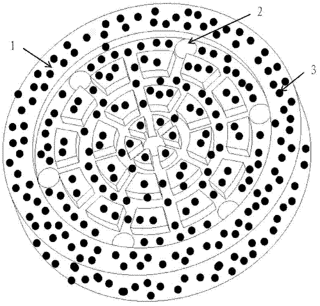
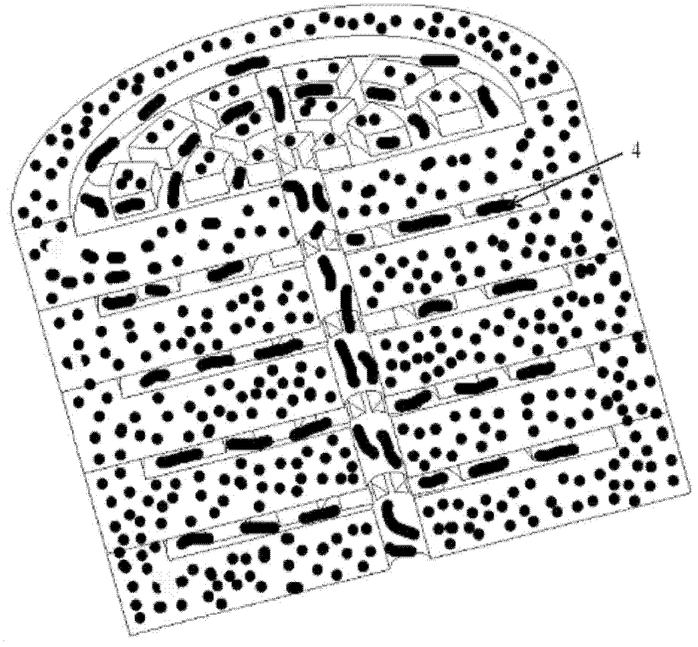
[0019] 1) According to the microstructural unit characteristics of liver, heart, kidney or lung, use 3D CAD software Pro / Engineer or UG to design a hydrogel unit model with bionic blood or culture fluid channel system, or use AutoCAD, CorelDraw and other software Design the photocuring mask of the bionic blood or culture fluid channel system. The hydrogel unit model channel system has a width of 50μm-1mm and a depth of 0.3-0.8mm.

[0020] 2) Export the designed hydrogel unit model in .STL format, use photocuring rapid prototyping or photolithography technology to manufacture the physical prototype of the hydrogel unit, and then use liquid medical silica gel or agarose with a mass fraction of 5%-10% The aqueous solution is...


PUM


Abstract

The invention provides a solid forming method of an active cell-hydrogel organ structure. The method comprises the following steps: firstly designing a hydrogel unit model according to natural organs; manufacturing a physical prototype of the unit by utilizing a micro-manufacturing technology, and then filling the physical prototype to obtain a negative structure; treating the surface of the negative structure, and then filling a negative die with a biological material-cell mixed solution; gelating the mixed solution in a cross-linking manner to obtain an active cell-hydrogel unit with a biomimetic blood flow channel; locating and superposing layer by layer and then performing secondary cross-linking to form the active cell-hydrogel organ structure; and finally culturing in vitro or implanting in vivo to construct a pre-vascularized active organ precursor. In the method, by assembling the two-dimensional active cell-hydrogel structure unit in a superposition manner, a complicated three-dimensional (3D) organ micro-fluidic channel system is constructed and cells are uniformly and sequentially seeded in a 3D space.

Description

technical field [0001] The invention relates to a forming method of an artificial active organoid structure, in particular to a three-dimensional forming method of an active cell-hydrogel organoid structure. Background technique [0002] In the 21st century, the in vitro manufacturing of human tissues and organs through the combination of manufacturing technology and biomedicine will bring fundamental changes to clinical treatment methods, thus benefiting thousands of patients. At present, artificial skin, cartilage, bone, bladder, etc. have entered the clinical application or experimental stage, but the in vitro manufacturing of important organs such as liver, kidney, lung, and heart has not made breakthroughs so far. The difficulty lies in the use of soft biomaterials to biomimetically construct a three-dimensional microstructure system and cell growth environment similar to natural organs in vitro. The traditional tissue engineering model of first forming the scaffold st...

Claims


Application Information

Patent Timeline
Patent Type & Authority Applications(China)
IPC IPC(8): A61F2/02
Inventor 贺健康李涤尘刘亚雄李骁连芩高琨卢秉恒
Owner XI AN JIAOTONG UNIV
Learn More