
A method for microRNA extraction from large-volume urine samples based on ultrafiltration concentration
An ultrafiltration concentration and large-volume technology, which is applied in the field of microRNA extraction, can solve the problems of low miRNA content, the inability to meet the experimental requirements of large-scale screening or expression detection, etc., to reduce the sample volume, shorten the time of adsorption and capture, and overcome the intractable effect
Patent Information
- Authority / Receiving Office
- CN · China
- Patent Type
- Applications(China)
- Current Assignee / Owner
- Publication Date
- 2011-12-14
- Estimated Expiration
- Not applicable · inactive patent
Smart Images
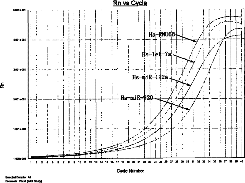
Figure 1 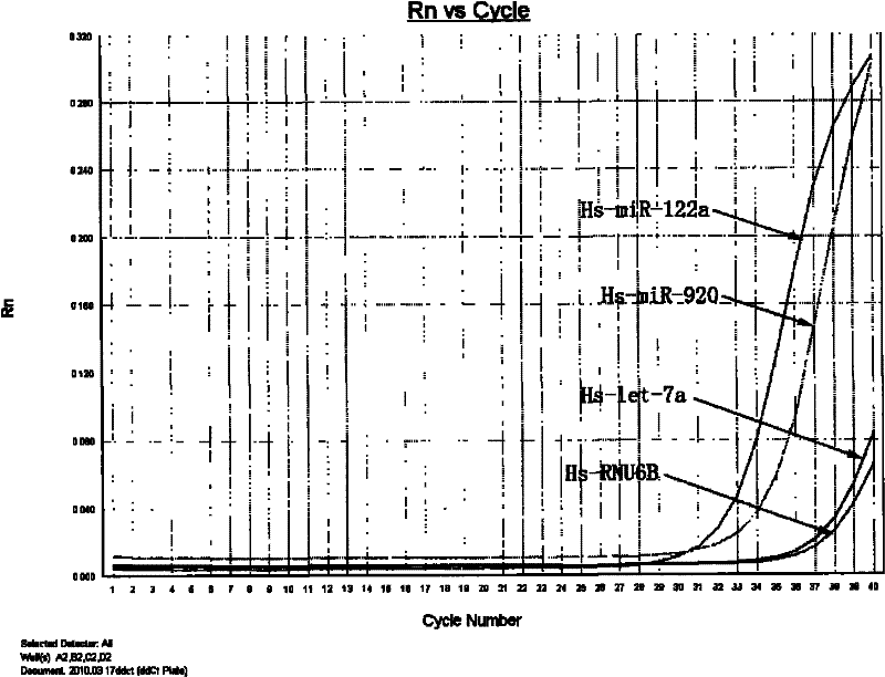
Figure 2 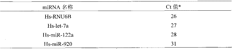
Figure 3
Abstract
Description
technical field
[0001] The invention relates to a method for extracting microRNA from large-volume urine samples. Background technique
[0002] MicroRNA (miRNA) is a 21-25nt long non-coding regulatory single-stranded small molecule RNA. Its main function is to regulate the expression of genes related to the growth, development and disease occurrence process of organisms. Existing studies have shown that the abnormal expression of some miRNAs is related to the occurrence and development of some diseases or cancers, and studies have found that miRNAs not only exist in tissue cells, but also exist stably in some body fluids such as serum, plasma and urine. Therefore, miRNAs are expected to It has become an important biomarker for detecting or predicting some diseases or tumors.
[0003] The commonly used technical methods for detecting miRNA are gene chips and fluorescent quantitative PCR, but the prerequisite for the application of these technical methods is to extract and pu...
Examples
Embodiment 1
[0014] The method of the present invention is used to extract free miRNA in the urine sample and detect its expression.
[0015] A urine sample is randomly obtained, and the health status of the sampling subject is unknown. The specific implementation process is as follows:
[0016] (1) Immediately add a certain amount of stabilizer to the urine sample (30ml), dissolve naturally and store at -80°C or proceed directly to the next experiment.
[0017] (2) Concentrate the urine sample to about 1 ml using an ultrafiltration tube (Amicon Ultra-15 3K, millipore), extract with Tris saturated phenol:chloroform (1:1), and take the supernatant. Then, the miRNA was extracted using a commercially available kit (miRNeasy Mini Kit, Qiagen).
[0018] (3) The obtained miRNA was reverse-transcribed using a reverse transcription kit (miScript Reverse Transcription Kit, Qiagen), and the reverse transcription conditions were: 37° C. for 60 min; 95° C. for 5 min.
[0019] (3) Fluorescence quanti...
Embodiment 2
[0025] Extraction of the same sample compared to the concentration method without ultrafiltration
[0026] Sample is identical with the sample of embodiment one, and specific implementation is as follows:
[0027] Refer to Example 1 for the pretreatment of the urine sample, without going through the ultrafiltration concentration step in Example 1, directly enter the Tris saturated phenol:chloroform (1:1) extraction in Step 3, and the remaining steps are the same as in Example 1. Since it is not concentrated by ultrafiltration, the amount of urine sample processed is limited, which is one-third of that in Example 1. The test results are shown in Table 2.
[0028] Table 2
[0029]
[0030] from Table 2 combined with the attached figure 2 It can be seen that after 40 cycles, the fluorescence value has not reached a stable value, and after converting to the Ct value, Hs-RNU6B and Hs-let-7a are not detected.