Method for detecting surface enhanced Raman scattering of cancer cells based on self-assembled material

A surface-enhanced Raman, self-assembly technology, applied in the field of analytical chemistry, to achieve the effect of homogeneous assembly structure

Active Publication Date: 2012-02-08
JIANGNAN UNIV
View PDF2 Cites 21 Cited by
  • Summary
  • Abstract
  • Description
  • Claims
  • Application Information

AI Technical Summary

Problems solved by technology

This enables single

Method used


Image

  • Method for detecting surface enhanced Raman scattering of cancer cells based on self-assembled material
  • Method for detecting surface enhanced Raman scattering of cancer cells based on self-assembled material
  • Method for detecting surface enhanced Raman scattering of cancer cells based on self-assembled material

Examples


Embodiment 1

[0056] Preparation of gold nanoparticles and gold nanorod probes

[0057] It is similar to the steps of preparing gold nanoparticles and gold nanorod probes in the patent applied by the applicant: a method for preparing self-assembled materials with surface-enhanced Raman activity, application number 101010605799.9, to prepare the gold nanoparticles required by this patent Probes for rods and gold nanoparticles.

[0058] 1) Preparation of gold nanoparticle probes

[0059] Add 95mL of water to a clean Erlenmeyer flask, then add 5mL of chloroauric acid with a concentration of 2g / L to the water, heat, boil, then add 2.5mL of trisodium citrate solution with a mass concentration of 1%, and stir while heating , the color of the solution changed from light yellow to red, and the reaction lasted for 6-8 minutes to allow the trisodium citrate to settle completely. Finally, the solution was cooled to room temperature, diluted to 100 mL, and stored at 4°C to obtain gold nanoparticles wi...

Embodiment 3

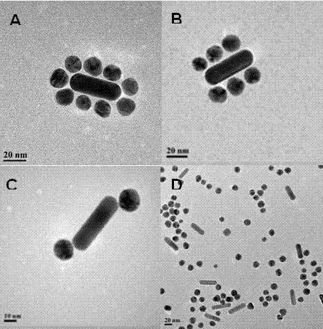
[0075] Example 3 Self-assembly of gold nanorod probes and gold nanoparticle probes

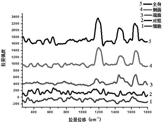
[0076] Gold nanoparticles assemble around gold nanorods to form satellite-like assembly structures

[0077] Take 2 μL of modified whole-body modified gold nanorods and 10 μL of modified gold nanoparticles, add 12 μL of 0.01M Tris-HCl with pH 7.5, 0.01% SDS, and 20 mM KCl hybridization buffer, mix well, and shake at room temperature. Shake the reaction for 12h.

[0078] Gold nanoparticles assemble around the sides of gold nanorods

[0079] Take 2 μL of modified side-modified and end-blocked gold nanorods and 9 μL of modified gold nanoparticles, add 11 μL of 0.01M Tris-HCl at pH 7.5, 0.01% SDS, and 20 mM KCl hybridization buffer, and mix well. The reaction was shaken at room temperature for 12h.

[0080] End-face assembly of gold nanoparticles and gold nanorods

[0081] Take 3 μL of modified gold nanorods with modified end faces and side-blocked sides and 3 μL of modified gold nanop...

Embodiment 4

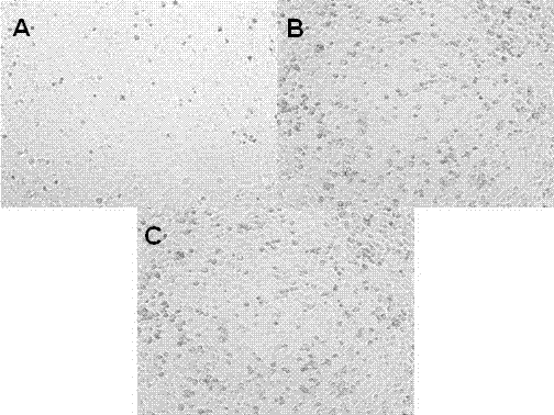
[0084] Example 4 Hela cell culture

[0085] The cancer cells used in this patent are Hela cells. Hela cell processing from flask to 96-well plate:

[0086] Remove the old medium in the culture flask, add 2 mL of pH 7.4 0.01M phosphate buffer to each culture flask, shake the culture flask slightly left and right, and then remove the phosphate buffer.

[0087] Then add 1mL of 0.25% trypsin digestion solution to each culture flask and digest for 3min.

[0088] The added tryptic solution was removed.

[0089] Add 2 mL of DMEM medium (provided by Shanghai Hufeng Biotechnology Co., Ltd.), and gently pipette several times until the cells on the wall of the culture flask are dispersed into the medium.

[0090] Add the above-prepared cell culture solution into the cell culture plate, and add 100 μL to each well.

[0091] Then the above cell culture plate was placed in the culture condition of 37°C, 5% CO 2 Cultivate in a constant temperature incubator for 12 h.


PUM

PropertyMeasurementUnit
Particle sizeaaaaaaaaaa

Abstract

The invention relates to a method for detecting surface enhanced Raman scattering of cancer cells based on a self-assembled material, and belongs to the technical field of analytical chemistry. The method provided by the present invention comprises: preparation of a gold nanorod probe and a gold nanoparticle probe, self-assembly of the gold nanorod probe and the gold nanoparticle probe, surface enhanced Raman detection of cancer cells. The surface enhanced Raman scattering (SERS) is a kind of Raman scattering, and can be briefly described as the following phenomenon: when a molecule is adsorbed on the especially-prepared metal surface, the Raman signal intensity is 105-106 times high than the expected value through simple calculation, such that the detection of the single molecule can be successful. With the present invention, the nano-material assembly is prepared and is applicable for detection of the cancer cells, wherein the prepared nano-material assembly has characteristics of uniform assembly structure, controllability and SERS activity.

Description

technical field [0001] The invention discloses a method for enhancing Raman detection on the surface of cancer cells based on self-assembled materials, which belongs to the technical field of analytical chemistry. Background technique [0002] Functional nanomaterials include quantum dots, gold nanoparticles, gold nanorods, etc. These nanomaterials have special optical, magnetic, electrical, and thermodynamic properties. The application of nanomaterials has become a current research hotspot. In particular, the special light absorption, light scattering effect and Raman enhancement effect of gold nanoparticles and gold nanorods make them the two most widely used materials in nanoscience and technology research as sensors. [0003] Monodispersed nanomaterials form some unique materials through self-assembly, which have properties such as plasmonic coupling, electromagnetic field enhancement, fluorescence quenching or enhancement, and multiple functional property coupling. The...

Claims


Application Information

Patent Timeline
IPC IPC(8): G01N21/65
Inventor 王利兵胥传来徐丽广匡华马伟严文静屈昌龙郝昌龙赵媛
Owner JIANGNAN UNIV
Why Eureka
Try Eureka