Radiological image detection apparatus, radiographic apparatus and radiographic system

A technology of radiographic images and radiography, which is applied in the evaluation of the musculoskeletal system, instruments for radiological diagnosis, diaphragms for radiological diagnosis, etc., can solve problems such as phase contrast image quality degradation, and achieve the effect of reducing scattering and reducing radiation

Inactive Publication Date: 2012-07-11
FUJIFILM CORP
View PDF4 Cites 40 Cited by
  • Summary
  • Abstract
  • Description
  • Claims
  • Application Information

AI Technical Summary

Problems solved by technology

However, such air bubbles may also degrade the quality of phase contrast images

Method used


Image

  • Radiological image detection apparatus, radiographic apparatus and radiographic system
  • Radiological image detection apparatus, radiographic apparatus and radiographic system
  • Radiological image detection apparatus, radiographic apparatus and radiographic system

Examples


Embodiment Construction

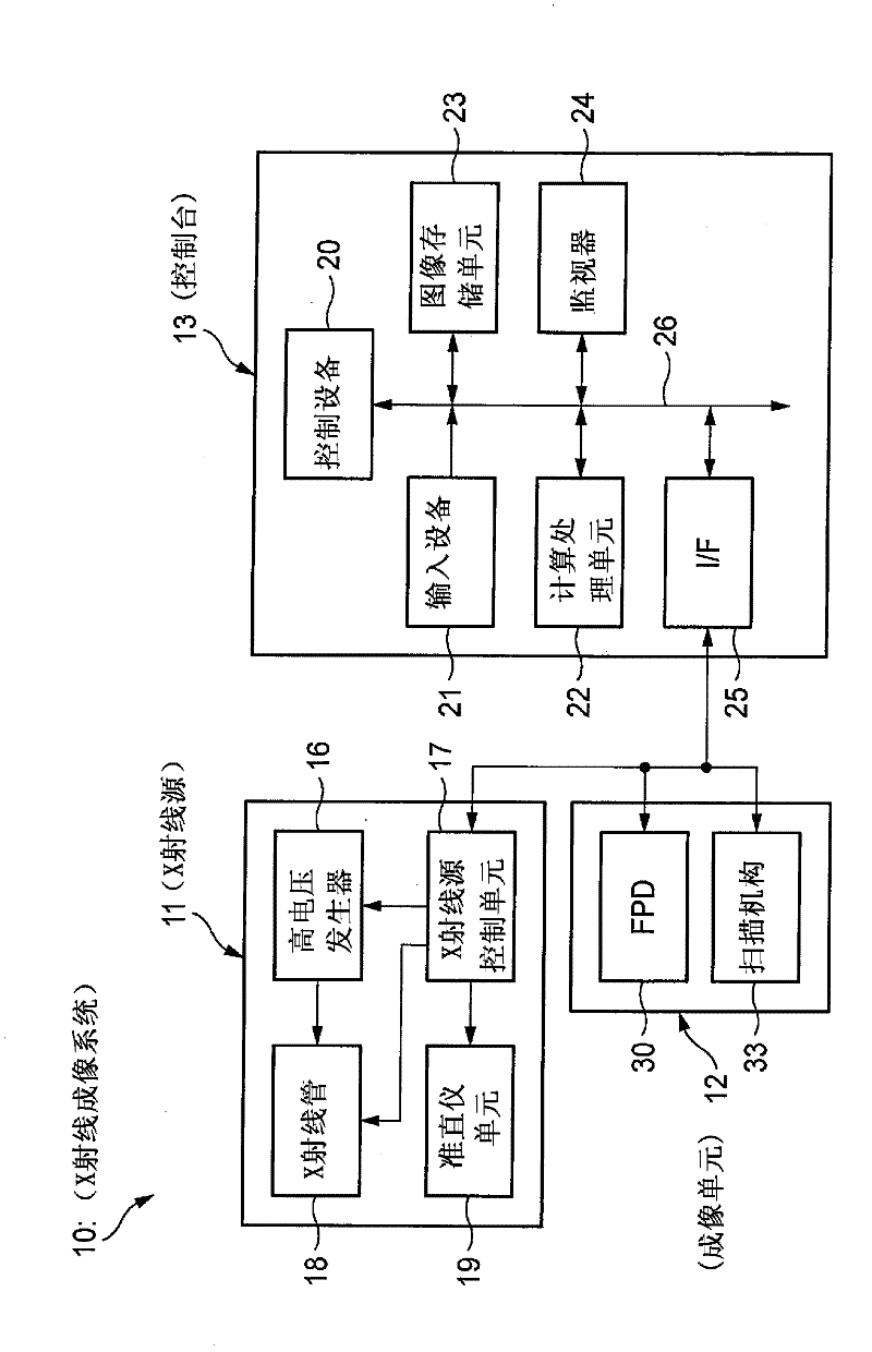
[0066] figure 1 An example of the configuration of a radiographic system for illustrating an illustrative embodiment of the present invention is shown, and figure 2 show figure 1 Control block diagram of the radiography system.

[0067] Meanwhile, the same configurations as those already described are denoted by the same reference numerals, and descriptions thereof are omitted. Differences from the already described configuration will be described.

[0068] The X-ray imaging system 10 is an X-ray diagnostic apparatus that performs imaging while a subject (patient) H is standing, and includes: an X-ray source 11 that irradiates the subject H; an imaging unit 12 that Used as a radiographic image detection device that is opposed to the X-ray source 11, wherein the subject H is interposed between the X-ray source 11 and the imaging unit, and that detects the transmitted light from the X-ray source 11 through the X-rays of the subject H, and thereby generate image data; and th...


PUM


Abstract

The invention discloses a radiological image detection apparatus, a radiographic apparatus and a radiographic system. The radiological image detection apparatus includes a first grating unit, a grating pattern unit, a radiological image detector, and an anti-scatter grating. The grating pattern unit has a period that substantially coincides with a pattern period of a radiological image formed by radiation having passed through the first grating unit. The radiological image detector detects the radiological image masked by the grating pattern unit. The anti-scatter grating is arranged on a path of the radiation incident onto the radiological image detector and removes scattered radiation. A smoothing process is performed for at least one of a surface and a backside of the anti-scatter grating intersecting with a traveling direction of the radiation.

Description

technical field [0001] The present invention relates to a radiographic image detection apparatus, a radiographic apparatus, and a radiographic system capable of enabling phase contrast imaging of a subject by using radiation such as X-rays. Background technique [0002] Since X-rays are attenuated according to the atomic number of elements constituting a material and the density and thickness of the material, X-rays are used as a probe for seeing through the inside of a subject. Imaging using X-rays is widely used in fields such as medical diagnosis and nondestructive testing. [0003] In a general X-ray imaging system, a subject is arranged between an X-ray source that irradiates X-rays and an X-ray image detector that detects the X-rays, and a transmission image of the subject is captured. In this case, the X-rays irradiated from the X-ray source to the X-ray image detector undergo attenuation (absorption ), and then incident on each pixel of the X-ray image detector. A...

Claims


Application Information

Patent Timeline
Patent Type & Authority Applications(China)
IPC IPC(8): A61B6/00
CPCA61B6/4233A61B6/06A61B5/45A61B6/4464A61B6/4452A61B6/484A61B6/4035A61B6/4021A61B6/502A61B6/4291
Inventor 多田拓司
Owner FUJIFILM CORP
Why Eureka
Try Eureka