Method and device for establishing three-dimensional craniomaxillofacial morphology model

A technology of three-dimensional topography and construction method, applied in the field of medical image generation and application, can solve the problems of inability to realize three-dimensional display, lack of three-dimensional topography simulation model, unable to faithfully reflect facial color and texture information, etc., and achieve high clinical value. And economic benefits, easy to promote, strong immersion effect

Inactive Publication Date: 2012-09-12
SHANGHAI NINTH PEOPLES HOSPITAL AFFILIATED TO SHANGHAI JIAO TONG UNIV SCHOOL OF MEDICINE
View PDF2 Cites 19 Cited by
  • Summary
  • Abstract
  • Description
  • Claims
  • Application Information

AI Technical Summary

Problems solved by technology

[0003] The technical problem to be solved by the present invention is to provide a method and device for constructing a three-dimensional topography model of the craniomaxillofacial region, which solves the problem that the prior art can only construct gray images of obj...

Method used


Image

  • Method and device for establishing three-dimensional craniomaxillofacial morphology model
  • Method and device for establishing three-dimensional craniomaxillofacial morphology model
  • Method and device for establishing three-dimensional craniomaxillofacial morphology model

Examples


Embodiment Construction

[0033] The specific implementation of the method for constructing the craniomaxillofacial three-dimensional topography model provided by the present invention will be described in detail below in conjunction with the accompanying drawings.

[0034] attached figure 1 Shown is a schematic diagram of the steps of the method for constructing a craniomaxillofacial three-dimensional topography model according to the present invention, and then the attached figure 1 The steps shown are described in detail.

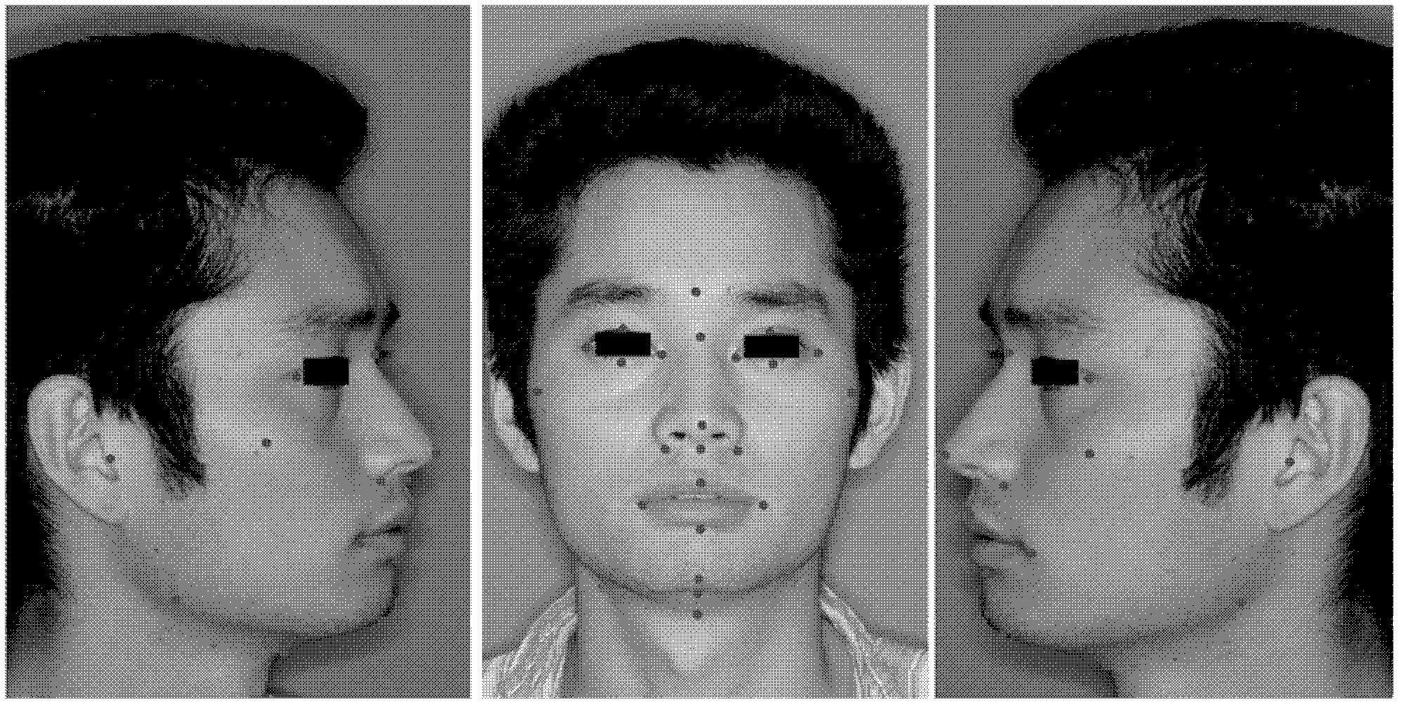
[0035] S11: Computed tomography was used to scan the cranio-maxillofacial face to obtain computed tomography data, and the front and side photos of the face were obtained through facial photography.

[0036] CT (Computer-aided Tomograph, computerized tomography) is a scanning method that uses computer technology to reconstruct the tomographic image of the measured object to obtain a three-dimensional tomographic image. CT plain scan refers to a continuous, non-interval, non-ove...


PUM


Abstract

A method for establishing a three-dimensional craniomaxillofacial morphology model includes the steps: (1) plain scanning craniomaxillofacial morphology by means of computerized tomography to acquire computerized tomography data, and performing facial photography to obtain facial anteroposterior and lateral photos; (2) inputting the computerized tomography data to a software system, three-dimensionally reconstructing facial soft tissues and transforming the facial soft tissues into a triangular facet model; (3) selecting anatomical mark points of the facial soft tissues; and (4) establishing texture mapping among the facial anteroposterior and lateral photos and the triangular facet model by the aid of the anatomical mark points of the facial soft tissues selected in the step (3) to establish the three-dimensional craniomaxillofacial morphology model. The software system has the function of picking facial features by the aid of mice, realizes mouse interaction operation for remarkable facial features, and picks facial feature points and image feature points in optional modes.

Description

technical field [0001] The invention relates to the field of medical image generation and application, in particular to a method and device for constructing a three-dimensional craniofacial topography model. Background technique [0002] The establishment and morphological analysis of the three-dimensional soft and hard tissue models of the maxillofacial region are of great significance to the study of facial growth, the diagnosis of facial deformities, the prediction of postoperative facial shape, and the evaluation of curative effects. At present, the methods for obtaining 3D image information of facial soft and hard tissues include moiré, 3D laser scanning, structured light technology, 3D stereo photography, grating projection measurement, CT, MRI, etc. Technologies such as Moiré moiré, structured light technology, laser scanning, and three-dimensional photography can only record the surface characteristics of objects, but are powerless to the internal structure of object...

Claims


Application Information

Patent Timeline
IPC IPC(8): G06T17/00
Inventor 于洪波沈国芳刘炳凯沈舜尧
Owner SHANGHAI NINTH PEOPLES HOSPITAL AFFILIATED TO SHANGHAI JIAO TONG UNIV SCHOOL OF MEDICINE
Why Eureka
Try Eureka