Method for staining actin filaments of suspension cells

A technique for actin filaments and suspension cells, which is applied in the preparation of test samples, etc., can solve the problems of non-adherent cells staining method operation, and suspension cells are difficult to adhere to and grow.

Inactive Publication Date: 2013-04-24
SHANGHAI OCEAN UNIV
View PDF3 Cites 0 Cited by
  • Summary
  • Abstract
  • Description
  • Claims
  • Application Information

AI Technical Summary

Problems solved by technology

[0005] Since it is difficult for suspension cells to adhere to the wall and grow in the culture process, it cannot be operated according to the staining method of adherent cells.

Method used


Image

  • Method for staining actin filaments of suspension cells
  • Method for staining actin filaments of suspension cells
  • Method for staining actin filaments of suspension cells

Examples


Embodiment 1

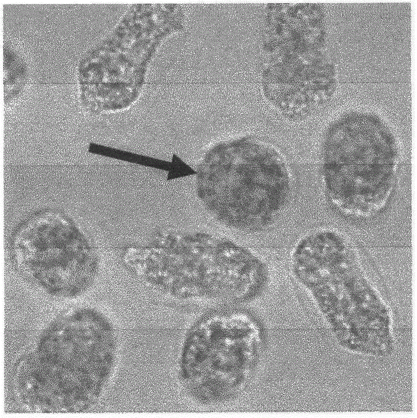
[0015] In this example, the actin filaments of the spores of Viola variegata were stained through the following steps. The specific steps are to collect single spores by centrifugation, and centrifuge at 800 rpm to collect suspension cells to be stained. Microfilament buffer (100mM PIPES, 10mM EGTA, 5mMMgSO 4 and 0.3M mannitol, pH 6.9) dilute the fluorescent marker substance AlexaFluor 488phalloidin (Molecular Probes, USA) of actin filaments to 5UmL -1 Prepare actin filament staining solution, and add 2% (v / v) glycerol to enhance the staining effect. The collected cells were suspended in the above staining solution, stained at room temperature and in the dark for 10 minutes, and centrifuged again at 800 rpm to collect the cells.

Embodiment 2

[0017] In this example, the actin filaments of the spores of Viola variegata were stained through the following steps. The specific steps are to collect single spores by centrifugation, and centrifuge at 900 rpm to collect suspension cells to be stained. Microfilament buffer (100mM PIPES, 10mM EGTA, 5mMMgSO 4 and 0.3M mannitol, pH 6.9) to dilute the fluorescent marker substance Alexa Fluor 488 phalloidin (Molecular Probes, USA) of actin filaments to 5UmL -1 Prepare actin filament staining solution, and add 2% (v / v) glycerol to enhance the staining effect. The collected cells were suspended in the above staining solution, stained at room temperature and in the dark for 10 minutes, and centrifuged again at 900 rpm to collect the cells.

Embodiment 3

[0019] In this example, the actin filaments of the spores of Viola variegata were stained through the following steps. The specific steps are to collect the single spores by centrifugation, and collect the suspension cells to be stained by centrifugation under the condition of 1000 rpm. Microfilament buffer (100mM PIPES, 10mM EGTA, 5mMMgSO 4 and 0.3M mannitol, pH 6.9) dilute the fluorescent marker substance AlexFlour 488phalloidin (Molecular Probes, USA) of actin filaments to 5UmL -1 Prepare actin filament staining solution, and add 2% (v / v) glycerol to enhance the staining effect. The collected cells were suspended in the above staining solution, stained at room temperature and in the dark for 10 minutes, and centrifuged again at 1000 rpm to collect the cells.

[0020] The cells collected in Example 2 were observed. In order to reduce the background caused by fluorescent substances in the observation, the collected cells could be centrifuged and washed once again with phosp...


PUM


Abstract

The invention relates to a method for staining actin filaments of suspension cells. The method comprises the following steps of: (A) collecting the suspension cells (single spores of porphyra yezoensis) to be stained by centrifuging under the condition of 800-1,000rpm (revolutions per minute); (B) diluting fluorescence-labeled substances Alexa Fluor 488phalloidin of the actin filaments by use of a microfilamentn buffering solution to a volume of 5UmL<-1>, and adding 2% by volume of glycerol for improving the staining effect to prepare a staining solution, wherein the microfilamentn buffering solution is prepared from 100mM PIPES, 10mM EGTA, 5mM MgSO4 and 0.3M mannitol, and the pH value of the microfilamentn buffering solution is 6.9; and (C) suspending the collected suspension cells into the staining solution, staining for 10 minutes at room temperature under the dark condition, and recollecting suspension cells by centrifuging under the condition of 800-1,000rpm (revolutions per minute). According to the method, the actin filaments of the single spores can be labeled well, and the cell debris and the excess staining solution can be removed in the process of centrifuging, so that the observation result is clearer.

Description

technical field [0001] The invention relates to the field of biomedicine, in particular to a method for staining actin filaments of suspended cells. Background technique [0002] Actin filament (actin filament) is a filament formed by the helical polymerization of actin molecules, which is one of the main components of the cytoskeleton. Actin filament, its association protion, and myosin constitute a chemical-mechanical system that utilizes chemical energy to generate mechanical motion, and has many effects on cell attachment, spreading, movement, endocytosis, and cell division. important role in cellular function. In the life activities of higher animals, the directional movement of cells is closely related to embryonic development, wound healing, immune response, tissue development and other activities. In addition, many major human diseases, such as tumorigenesis and metastasis, are also closely related to cell movement. Therefore, in order to intuitively observe the s...

Claims


Application Information

Patent Timeline
Patent Type & Authority Applications(China)
IPC IPC(8): G01N1/30
Inventor 李琳严兴洪
Owner SHANGHAI OCEAN UNIV
Why Eureka
Try Eureka