VISTA regulatory T cell mediator protein, VISTA binding agents and use thereof

A protein and cell technology, used in peptide/protein components, anti-inflammatory agents, fusion polypeptides, etc., can solve problems such as insufficient control of inflammation and damage

Active Publication Date: 2013-05-22
TRUSTEES OF DARTMOUTH COLLEGE THE
View PDF33 Cites 23 Cited by
  • Summary
  • Abstract
  • Description
  • Claims
  • Application Information

AI Technical Summary

Problems solved by technology

B7.1 and B7.2 knockout (KO) mice are impaired in the adaptive immune response, whereas CTLA-4

Method used


Image

  • VISTA regulatory T cell mediator protein, VISTA binding agents and use thereof
  • VISTA regulatory T cell mediator protein, VISTA binding agents and use thereof
  • VISTA regulatory T cell mediator protein, VISTA binding agents and use thereof

Examples


Embodiment 1

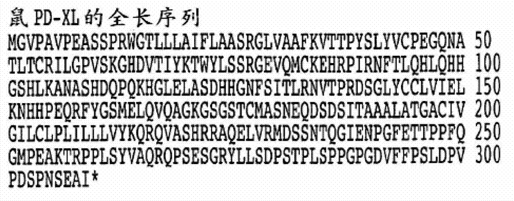
[0364] Example 1: Cloning and sequence analysis of PD-L3OR VISTA.

[0365] PD-L3OR VISTA and Treg-sTNF were identified by the overall transcription profile analysis of resting Treg, αCD3 activated Treg and αCD3 / αGITR activated Treg. ΑGITR was chosen for this analysis because the triggering of GITR on Treg has been shown to suppress their contact-dependent inhibitory activity (Shimizu, et al. (2002) supra). in PD-L3OR VISTA and Treg-sTNF were identified on the DNA array based on their unique expression patterns (Table 1). PD-L3OR VISTA showed increased expression in αCD3-activated Treg, and decreased expression in the presence of αGITR; and Treg-sTNF showed αCD3 / αGITR-dependent enhancement of expression.

[0366] Purified CD4+CD25+T cells were stimulated in overnight culture with no, αCD3 or αCD3 / αGITR, and RNA was isolated for real-time PCR analysis. The listed expressions are compared to actin.

[0367] Table 1

[0368]

[0369] The Affymetrix analysis of activated and resting CD...

Embodiment 2

[0374] Example 2: PD-L3ORVISTA expression study by RT-PCR analysis and flow cytometry

[0375] As shown in the experiment in Figure 3, RT-PCR analysis was used to determine the mRNA expression pattern of PD-L3OR VISTA in mouse tissues ( Figure 3A ). PD-L3OR VISTA is mainly expressed on hematopoietic tissues (spleen, thymus, bone marrow) or tissues with abundant lymphocyte infiltration (ie, lung). Weak expression was also detected in non-hematopoietic tissues (ie, heart, kidney, brain, and ovary). Analysis of several hematopoietic cell types revealed that PD-L3OR VISTA is expressed on peritoneal macrophages, splenic CD11b+ monocytes, CD11c+DC, CD4+ T cells and CD8+ T cells, but the expression level is low on B cells ( Figure 3B ). This expression pattern is also highly consistent with the GNF (Genomics Institute of Novartis Research Foundation) gene array database (Su et al., (2002), Proc Natl Acad Sci USA 99, 4465-4470) and the NCBIGEO (gene expression omnibus) database (Figur...

Embodiment 3

[0383] Example 3: The functional effect of PD-L3OR VISTA signal transduction on CD4+ and CD8+ T cell responses

[0384] The PD-L3OR VISTA-Ig fusion protein was produced to study the regulation of PD-L3OR VISTA on CD4+ T cell response. The PD-L3OR VISTA-Ig fusion protein contains the extracellular domain of PD-L3OR VISTA fused to the Fc region of human IgG1. When immobilized on a microplate, PD-L3OR VISTA-Ig, but not control Ig, inhibited the proliferation of crudely purified CD4+ and CD8+ T cells in response to anti-CD3 stimulation bound to the plate, as determined by inhibited cell division ( Figure 9A -B). The PD-L3OR VISTA Ig fusion protein does not affect the absorption of anti-CD3 antibodies into the plastic wells, as determined by ELISA (data not shown), thereby eliminating the possibility of non-specific inhibition. PD-1KO CD4+ T cells are also suppressed ( Figure 9C ), indicating that PD-1 is not a receptor for PD-L3OR VISTA. The inhibitory effects of PD-L1-Ig and PD-...


PUM


Abstract

The present invention relates to a novel regulatory T cell protein. This protein, designated PD-L3 OR VISTA resembles members of the PD-L1 family, identified a novel and structurally-distinct, Ig-superfamily inhibitory ligand, whose extracellular domain bears homology to the B7 family ligand PD-L1. This molecule is designated as PD-L3 OR VISTA or V-domain Immunoglobulin Suppressor of T cell Activation (VISTA). Expression of VISTA is primarily within the hematopoietic compartment and is highly regulated on myeloid APCs and T cells. Therapeutic intervention of the VISTA inhibitory pathway represents a novel approach to modulate T cell-mediated immunity for the treatment of a wide variety of cancers, e.g., ovarian, bladder cancer and melanomas. Also, VISTA proteins, especially multimeric VISTA proteins and antibodies may be used to suppress T cell immunity in autoimmune disease, allergy, infection and inflammatory conditions, e.g. multiple sclerosis and artritic conditions such as RA.

Description

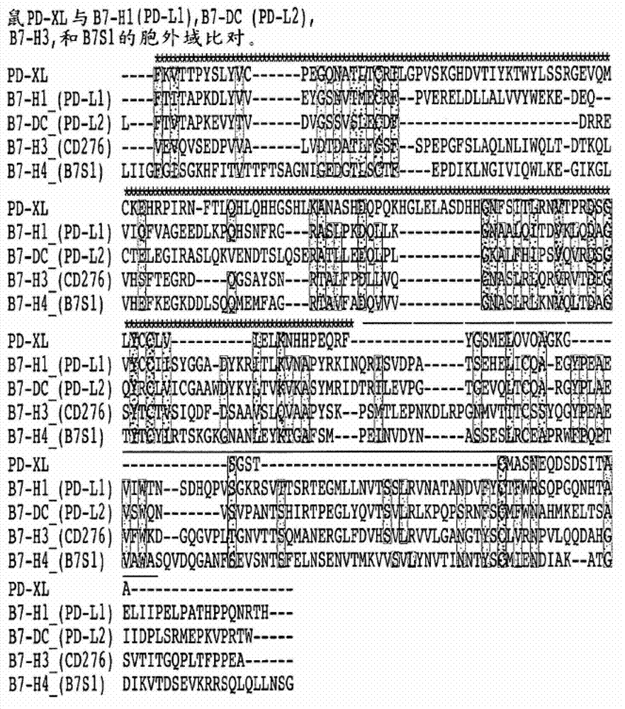
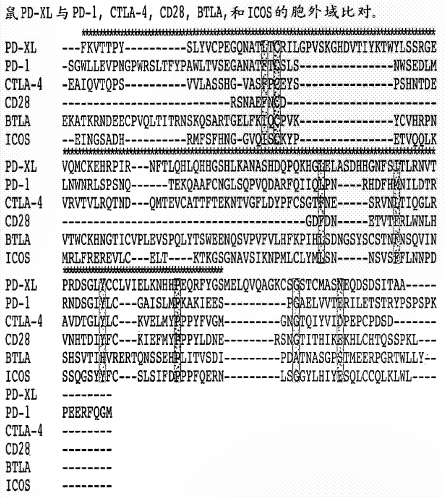
[0001] Introduction [0002] This patent application requires U.S. Patent Serial No. 12 / 732,371 filed on March 26, 2010, U.S. Provisional Application No. 61 / 390,434 filed on October 6, 2010, and U.S. Provisional Application No. filed on January 26, 2011 61 / 436,379 and U.S. Provisional Application No. 61 / 449,882 filed on March 7, 2011. All these patent applications are incorporated herein in their entirety by application. [0003] This patent application relates to the novel, hematopoietic restricted, structurally different Ig superfamily inhibitory ligands that we have discovered, characterized and determined, which are called T cell activated V-region containing immunoglobulin inhibitors (VISTA) or PD -L3. Its extracellular domain has homology with the B7 family ligand PD-L1, and like PD-L1, VISTA has a profound impact on immunity. However, unlike PD-L1, the expression of VISTA is only located in the hematopoietic chamber. Expression is most common in bone marrow antigen presen...

Claims


Application Information

Patent Timeline
IPC IPC(8): C07K14/47C07K19/00C07K16/18A61K38/17A61K39/395A61P37/00A61P19/02A61K39/00
CPCC07K16/2803C07K14/70503C07K16/28A61K2039/505C07K2317/76C07K2319/30A61P1/02A61P1/04A61P1/14A61P1/16A61P1/18A61P11/00A61P11/02A61P11/06A61P11/08A61P11/16A61P13/02A61P13/12A61P15/08A61P15/10A61P17/00A61P17/02A61P17/04A61P17/06A61P17/14A61P19/02A61P19/06A61P21/00A61P21/02A61P21/04A61P25/00A61P25/06A61P25/08A61P25/28A61P27/02A61P27/16A61P29/00A61P31/00A61P31/04A61P31/06A61P31/10A61P31/12A61P31/14A61P31/18A61P31/20A61P33/06A61P33/10A61P33/12A61P35/00A61P35/02A61P37/00A61P37/02A61P37/06A61P37/08A61P39/02A61P43/00A61P5/00A61P5/14A61P5/38A61P7/00A61P7/04A61P7/06A61P9/00A61P9/06A61P9/08A61P9/10A61P3/10Y02A50/30A61K39/395C07K14/47C07K19/00C07K16/18A61K38/17
Inventor R·J·诺雷L·王
Owner TRUSTEES OF DARTMOUTH COLLEGE THE
Why Eureka
Try Eureka