Method for determining effectiveness of medicine containing antibody as component

A determination method and a technology for pharmaceuticals, applied in the field of tissue staining, can solve problems such as low effect, and achieve the effect of promoting cancer treatment

Inactive Publication Date: 2013-06-12
TOHOKU UNIV
View PDF7 Cites 3 Cited by
  • Summary
  • Abstract
  • Description
  • Claims
  • Application Information

AI Technical Summary

Problems solved by technology

[0006] In addition, the effective rate of trastuzumab single-drug administration in breast cancer patients is 15 to 34%, and its low effect as an antibody drug is also a problem

Method used


Image

  • Method for determining effectiveness of medicine containing antibody as component
  • Method for determining effectiveness of medicine containing antibody as component
  • Method for determining effectiveness of medicine containing antibody as component

Examples


Embodiment 1

[0090] Method for preparing tissue sample immunostained using IHC-QDs method

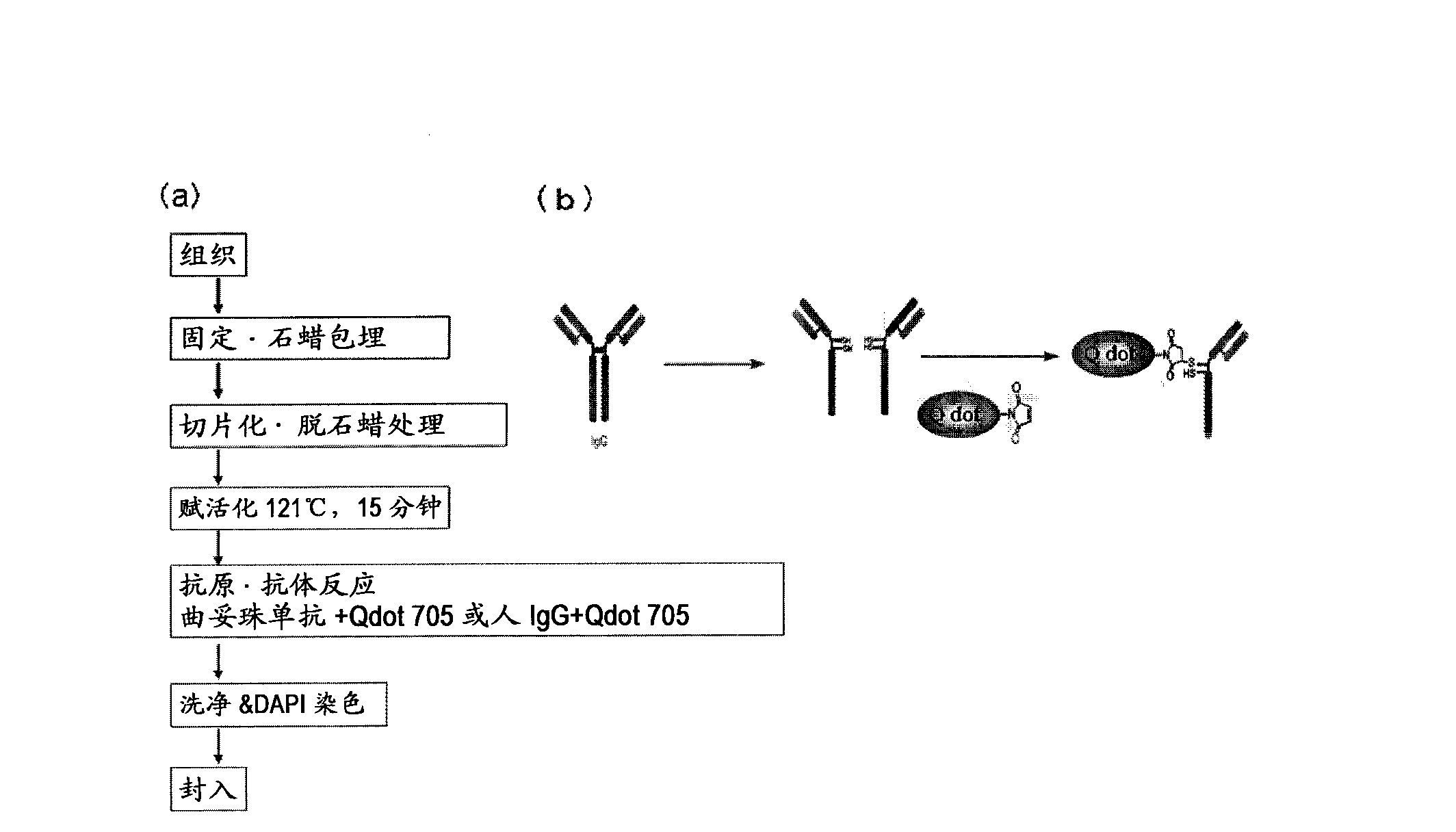
[0091]As human breast cancer pathological tissues, 6 cases with a score of 0, 6 cases with a score of 1, 11 cases with a score of 2, and 14 cases with a score of 3 based on the IHC-DAB method were selected, totaling 37 cases. In addition, regarding the background of the case, as shown in Table 1, it is considered that there is no bias. With respect to the tissues of the above 37 cases, tissue samples were prepared according to the method used in usual histopathological diagnosis. That is, the mammary tissue test sample of a cancer patient is fixed in formalin, dehydrated with ethanol, then processed with xylene, immersed in high-temperature paraffin and embedded in paraffin, and the tissue sample ( figure 1 (a)). Next, the above-mentioned tissue samples were sectioned at 2 to 4 μm, deparaffinized with xylene, treated with ethanol, and washed with deionized water. In order for the antibody to effi...

Embodiment 2

[0097] Problems with fluorescent immunohistostaining

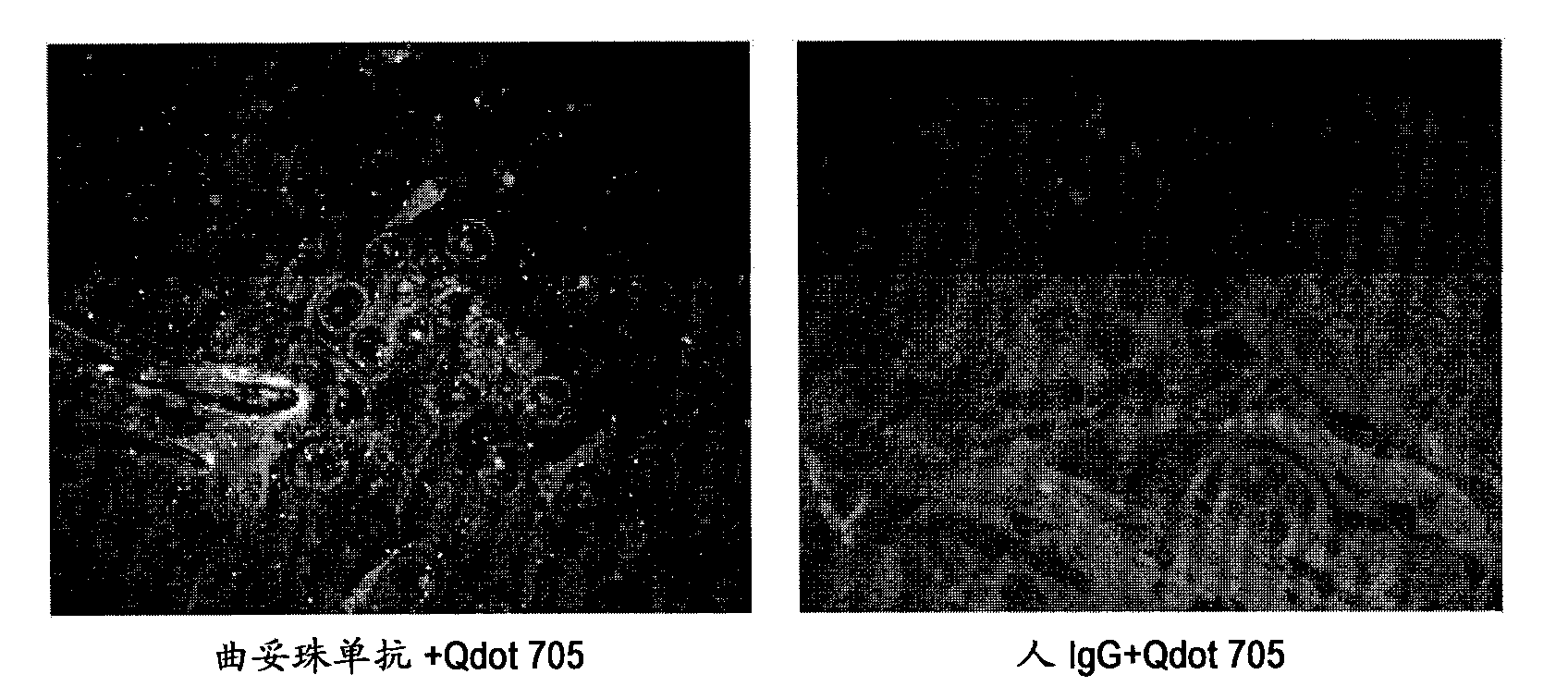
[0098] For the immunostained tissue sample prepared in Example 1 above, a combination of a confocal unit (manufactured by Yokogawa Electric Corporation), a fluorescence microscope (manufactured by Olympus Corporation), and an electron multiplier CCD (EM-CCD) imaging device (Andor (manufactured by the company) was irradiated with excitation light at 488nm, and then a fluorescence image (fluorescence still image) of quantum dot fluorescent particles (labeled to trastuzumab) was obtained using a bandpass filter with a wavelength acquisition range of 695 to 740nm ). figure 2 In the case of the case evaluated as score 3 by the IHC-DAB method as an example, it was shown that the fluorescence of quantum dot fluorescent particles was hardly observed when human IgG+Qdot705 was used as a control, but in contrast, trastuzumab+Qdot705 was used The fluorescence of quantum dot fluorescent particles was observed at Qdot705. These resu...

Embodiment 3

[0101] Creation of corrected fluorescence images with autofluorescence removed

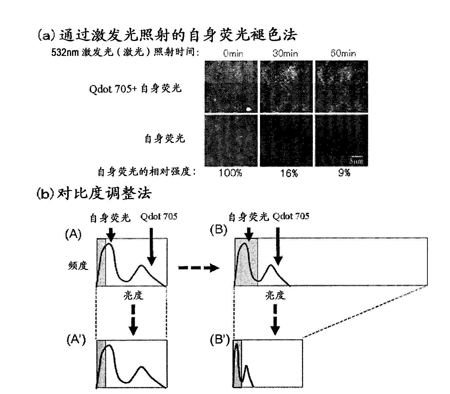
[0102] Fluorescence still images with zero (0) fluorescence intensity of autofluorescence as background are required. In this way, the total fluorescence in the fluorescence still image can be calculated as the fluorescence derived from the quantum dot fluorescent particles. The contrast adjustment method of the above-mentioned embodiment 2 is a division method of fluorescence intensity, and zero (0) cannot be obtained by division, so it must be an image processing method using a subtraction method that can obtain zero (0). Thus, as an image processing method for removing autofluorescence in a fluorescent still image, the method described below is considered. First, the breast cancer tissue sample immunostained with quantum dot fluorescent particles is irradiated with excitation light (laser) with an excitation wavelength of 488 nm, and a fluorescence image of quantum dot fluorescent particles is...


PUM

PropertyMeasurementUnit
molecular weightaaaaaaaaaa

Abstract

Provided is a method for determining effectiveness of medicine containing, as components, antibodies that are active components of an antibody preparation such as trastuzumab, or antibodies for a target to a target site of active component, by determining the quantity of the protein recognized by the antibodies with high accuracy, in a quantitative tissue staining method for biological tissue. The effectiveness of the medicine containing antibodies as components is determined by using the tissue staining method including the following steps: a step of labeling the antibodies in a medicine that contains antibodies as components with a fluorescent substance, and bringing the fluorescently labeled antibodies into contact with a tissue sample; a step of irradiating the tissue site contacted with the antibodies with excitation light to obtain a fluorescence image; a step of obtaining an intrinsic fluorescence image in the vicinity of the short-wavelength side or long-wavelength side of the acquisition area of fluorescence wavelength emitted from the fluorescent substance, in the same field of vision and in the same focus as those of the fluorescence image; a step of obtaining a collected fluorescence image by image-processing to eliminate the fluorescent intensity of the intrinsic fluorescence image from the fluorescent intensity of the fluorescence image; a step of counting the number of cells at the tissue site contacted with the antibodies; a step of measuring the average fluorescent intensity of one particle of fluorescent particles; and a step of calculating the number of fluorescent particles per cell.

Description

technical field [0001] The present invention relates to a method for determining the effectiveness of pharmaceuticals containing antibodies as components, using a tissue staining method that effectively eliminates the influence of autofluorescence and enables quantitative analysis with high accuracy. Background technique [0002] Cancer, together with vascular system diseases represented by myocardial infarction and cerebral infarction, are the two major causes of death in adults. For example, although the incidence rate of breast cancer in Japan is lower than that of European and American countries, it tends to increase in recent years. In 1998, it surpassed the incidence rate of gastric cancer and became the first female incidence rate. According to the statistics of the Ministry of Health, Labor and Welfare of Japan in 2005, which is the most recent report, the annual number of breast cancer patients exceeds 50,000. This number is also increasing year by year all over th...

Claims


Application Information

Patent Timeline
Patent Type & Authority Applications(China)
IPC IPC(8): G01N33/574G01N21/64G01N21/78G01N33/536
CPCG01N21/6428G01N21/6456G01N21/6486G01N33/582G01N33/6854G01N33/94G01N2800/52
Inventor 宫下穰权田幸祐大内宪明武田元博
Owner TOHOKU UNIV
Why Eureka
Try Eureka