Multi-photon fluoroscopy attachment module for surgical microscope

A technology for surgical microscopes and auxiliary modules, applied in the field of microscope systems, can solve problems such as high magnification, achieve the effects of no side effects, and reduce the number of waiting and interruptions

Inactive Publication Date: 2013-08-21
LEICA MICROSYSTEMS (SCHWEIZ) AG
View PDF7 Cites 2 Cited by
  • Summary
  • Abstract
  • Description
  • Claims
  • Application Information

AI Technical Summary

Problems solved by technology

[0004] However, especially for pathological evaluation of tissues, higher magnification is required

Method used


Image

  • Multi-photon fluoroscopy attachment module for surgical microscope

Examples


Embodiment Construction

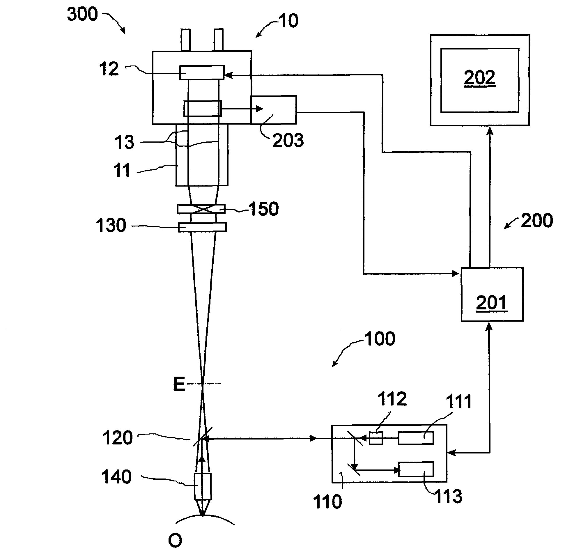
[0024] exist figure 1 In , a preferred microscope system is shown schematically and designated 300 as a whole. In a preferred embodiment, microscope system 300 includes surgical microscope 10 and inspection device 200 as a light microscope.

[0025] The examination device 200 includes an auxiliary module 100 . The auxiliary module 100 is placed in use in the main optical path of the surgical microscope 10 between the objective 11 and the object O to be observed. To this end, the auxiliary module can be attached to the surgical microscope 10 itself or to a support (not shown), on which the surgical microscope can also be mounted. In particular, the auxiliary module 100 is movably supported so that it can be inserted into the primary optical path as desired and removed therefrom after use.

[0026] The auxiliary module 100 has a multiphoton fluoroscope 110 . The multiphoton fluoroscope 110 comprises: a light source for emitting excitation light, here in the form of an infrar...


PUM


Abstract

The present invention relates to an attachment module (100) for a surgical microscope (10), the attachment module (100) being insertable into a main optical path between an objective (11) of the surgical microscope and an object (O) being observed, and including: a multi-photon fluoroscope (110) including a light source (111) for emitting excitation light, a scanning device (112) for directing the excitation light onto the object (O), and a detector (113) for detecting fluorescent light emitted from the object (O); and input coupling optics (120) for reflecting the excitation light from the scanning device onto the object (O).

Description

technical field [0001] The invention relates to an auxiliary module for a surgical microscope, an examination device comprising such an auxiliary module and a microscope system comprising such an examination device. Background technique [0002] In microscope-assisted surgery, light microscopic imaging can be supported by other methods. For example, it is known from the article "Combining Optical Coherence Tomography (OCT) with an Operating Microscope, E. Lankenau et al., Advances in Medical Engineering, Springer Verlag, Vol. 114, pp. 343-348" that an operating microscope can be equipped with an OCT ( Optical coherence tomography) unit so that images from deeper layers can be utilized in addition to light microscopic images of the eye, especially in ophthalmology. The magnification of the surgical microscope and the magnification of the OCT unit are of the same order of magnitude. [0003] In neurosurgery, as described, for example, in DE 10 2005 005 984 A1, fluorescence m...

Claims


Application Information

Patent Timeline
IPC IPC(8): A61B19/00G02B21/08G02B21/22G02B21/36
CPCH04N7/18G02B21/0012A61B19/5223G01N21/64G01N21/6458A61B19/52A61B90/20
Inventor 乌尔里克·桑德
Owner LEICA MICROSYSTEMS (SCHWEIZ) AG
Why Eureka
Try Eureka