Eureka AIR delivers breakthrough ideas for toughest innovation challenges, trusted by R&D personnel around the world.

Environmental scanning electron microscope detection method of a kind of hydration molecular form of polymer

An environmental scanning electron microscope and hydration molecule technology, which is applied in the direction of material analysis using measurement of secondary emissions, can solve the problem that the real shape of the polymer cannot be completely maintained, the real shape of the polymer cannot be observed, and the polymer structure collapses. and other problems, to achieve the effect of simple equipment and detection conditions

Inactive Publication Date: 2015-12-09
CHINA UNIV OF PETROLEUM (BEIJING)
View PDF2 Cites 0 Cited by
  • Summary
  • Abstract
  • Description
  • Claims
  • Application Information

AI Technical Summary

Problems solved by technology

[0004] 1) Observation with a conventional scanning electron microscope after direct drying: In this method, the polymer structure is severely damaged during the drying process, and the true morphology of the polymer in aqueous solution cannot be observed
[0005] 2) Observation with a conventional scanning electron microscope after freeze-drying: This method observes the sample after freeze-drying at low temperature (less than 0°C). The disadvantage of this method is that during the freeze-drying process, the polymer structure will change. collapsed, there is no way to fully maintain the true shape of the polymer
[0006] 3) Observation with an environmental scanning electron microscope with a cold stage: This method requires the purchase of a special cold stage for the electron microscope, which is expensive and needs to be installed on the electron microscope every time an experiment is performed, which is cumbersome to operate and prone to air leakage

Method used


Image

  • Environmental scanning electron microscope detection method of a kind of hydration molecular form of polymer
  • Environmental scanning electron microscope detection method of a kind of hydration molecular form of polymer
  • Environmental scanning electron microscope detection method of a kind of hydration molecular form of polymer

Examples


Embodiment 1

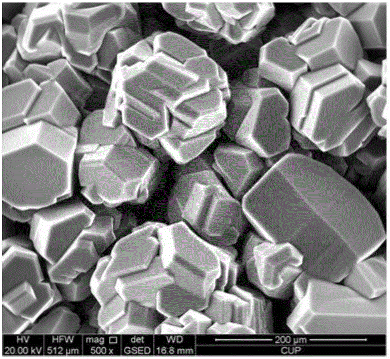
[0033] This example provides an environmental scanning electron microscope detection method for the hydration molecular morphology of the hydrophobic association polymer, which is to observe the microscopic morphology of the hydration molecular morphology of the hydrophobic association polymer AP-P4, the hydrophobic association polymer AP-P4 is an industrial product, and the manufacturer is Sichuan Guangya Co., Ltd. The mass fraction of the polymer is 1400 μg / g, and the balance is water.

[0034] The detection method is carried out according to the following steps:

[0035] (1) Put the copper block into the beaker, pour liquid nitrogen into the beaker to make the copper block cryogenic;

[0036] (2) Take out the copper block after 15 minutes, and drop a drop of hydrophobic association polymer sample on the copper block;

[0037] (3) Quickly transfer the copper block to the sample chamber of the environmental scanning electron microscope;

[0038] (4) Install the ring scan d...

Embodiment 2

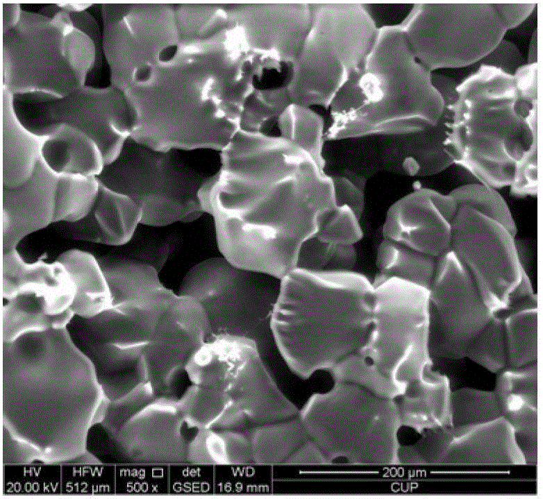
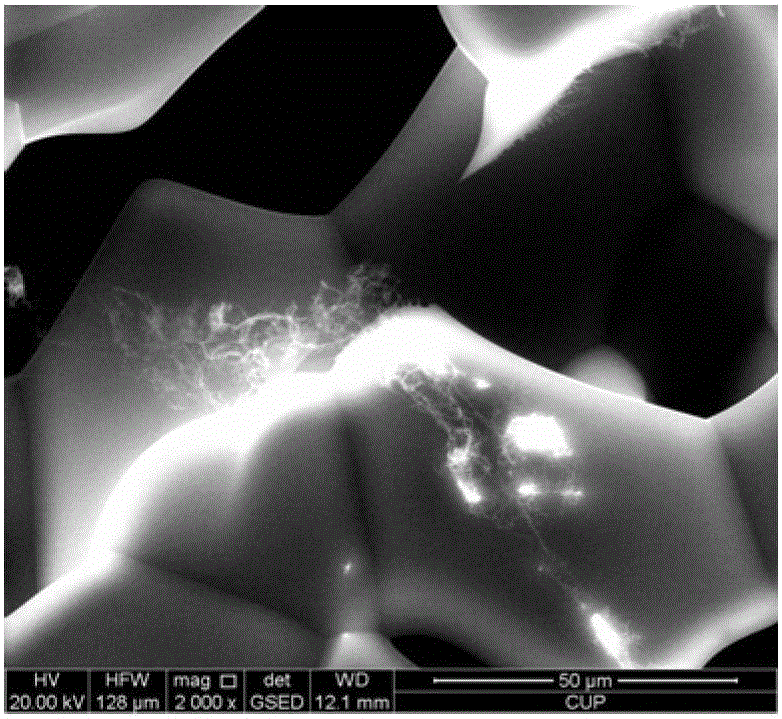
[0041] This embodiment provides an environmental scanning electron microscope detection method for the hydration molecular form of the gel, which is to observe the microscopic form of the hydration molecule of the gel. The gel is composed of 10wt% acrylic acid, propylene amide 23wt%, N,N-methylenebisacrylamide 0.2wt% and the balance of water.

[0042] The detection method is carried out according to the following steps:

[0043] (1) Put the copper block into the beaker, pour liquid nitrogen into the beaker to make the copper block cryogenic;

[0044] (2) Take out the copper block after 15 minutes, and place the gel sample (block shape) with a diameter of 0.5-1cm on the copper block;

[0045] (3) Quickly transfer the copper block to the sample chamber of the environmental scanning electron microscope;

[0046] (4) Install the ring scan detector of the environmental scanning electron microscope, and adjust the pressure of the environmental scanning electron microscope to 267Pa...


PUM

PropertyMeasurementUnit
diameteraaaaaaaaaa
heightaaaaaaaaaa

Abstract

The invention relates to an environmental scanning electron microscope detection method for hydration molecular morphology of a polymer. The method comprises the following steps of: immersing a copper block in liquid nitrogen; taking out the copper block after 15min, and placing a sample of the water-containing polymer on the copper block; rapidly transferring the copper block into a sample chamber of an environmental scanning electron microscope; mounting a circular scanning detector of the environmental scanning electron microscope, and performing vacuum pumping to adjust the pressure of the environmental scanning electron microscope to 10-600Pa; and performing detection and observation on a microstructure of the sample of the water-containing polymer after the pressure reaches 10-600Pa. By adopting the environmental scanning electron microscope detection method for observing the hydration molecular morphology of the polymer, required equipment and detection conditions are relatively simple, the internal microstructure of the polymer, a cross-linked polymer, gel or other water-containing samples can be effectively observed; and the environmental scanning electron microscope detection method is a good detection method for the microscopic morphology of the sample of the water-containing polymer.

Description

technical field [0001] The invention relates to an environmental scanning electron microscope detection method of the hydration molecular form of a polymer, belonging to the technical field of material detection. Background technique [0002] Polymers, cross-linked polymers, gels, etc. are often used as oil displacement agents and profile control agents in oil fields to enhance oil recovery. Different polymers have different hydration molecular forms, and their usage methods and applicable occasions are also different. Therefore, polymer hydration molecular forms have always been a concern of polymer synthesis and application researchers. Due to the interfacial tension on the surface of the liquid sample, the appearance of the liquid sample under different magnifications is basically a plane. In fact, the observation of the liquid sample mainly lies in the observation of the solid substance in the liquid or the structural changes that can form a solid substance. . [0003]...

Claims


Application Information

Patent Timeline
Patent Type & Authority Patents(China)
IPC IPC(8): G01N23/22
Inventor 焦淑静郑晓宇陈海玲姜大强杨峰崔立山
Owner CHINA UNIV OF PETROLEUM (BEIJING)
Why Eureka
Eureka Blog
Learn More