General surgery three-dimensional micrography camera shooting presentation device

A demonstration device and surgical operation technology, applied in the fields of surgery, application, medical science, etc., can solve the problems of expensive, complex and large structures, and achieve the effect of avoiding discomfort and adverse reactions, and facilitating teaching and academic demonstrations

Active Publication Date: 2013-10-09
上海昌宁医疗科技有限公司
View PDF8 Cites 14 Cited by
  • Summary
  • Abstract
  • Description
  • Claims
  • Application Information

AI Technical Summary

Problems solved by technology

The structure of the whole device is compl

Method used


Image

  • General surgery three-dimensional micrography camera shooting presentation device
  • General surgery three-dimensional micrography camera shooting presentation device
  • General surgery three-dimensional micrography camera shooting presentation device

Examples


Embodiment Construction

[0018] The specific implementation manners of the present invention will be described in further detail below in conjunction with the accompanying drawings.

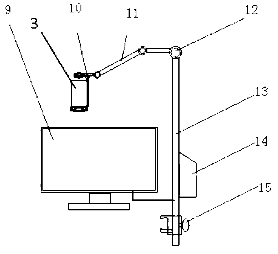
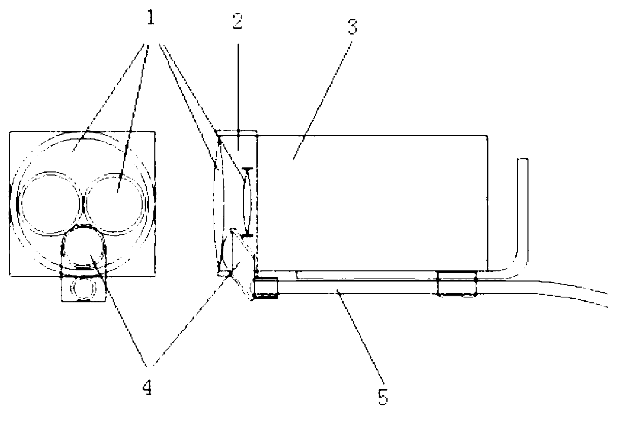
[0019] refer to figure 1 As shown, the adjusting bracket 13 of the present invention is a bedside multi-joint fixed bracket, which is provided with a pan-tilt bottom seat 8 and a pan-tilt 10 for fixing a 3D high-definition digital camera 3, a connecting rod 11, a universal adjustment knob 12, and a fixed operating bed. The knob 15 and the power supply 14 of the 3D high-definition digital camera 3 and the LED light source 7 can be installed on the adjustment bracket 13. The 3D high-definition digital camera 3 is fixed on the adjustment bracket 13 and connected with the 3D high-definition liquid crystal display 9 through a high-definition signal line (HDMI).

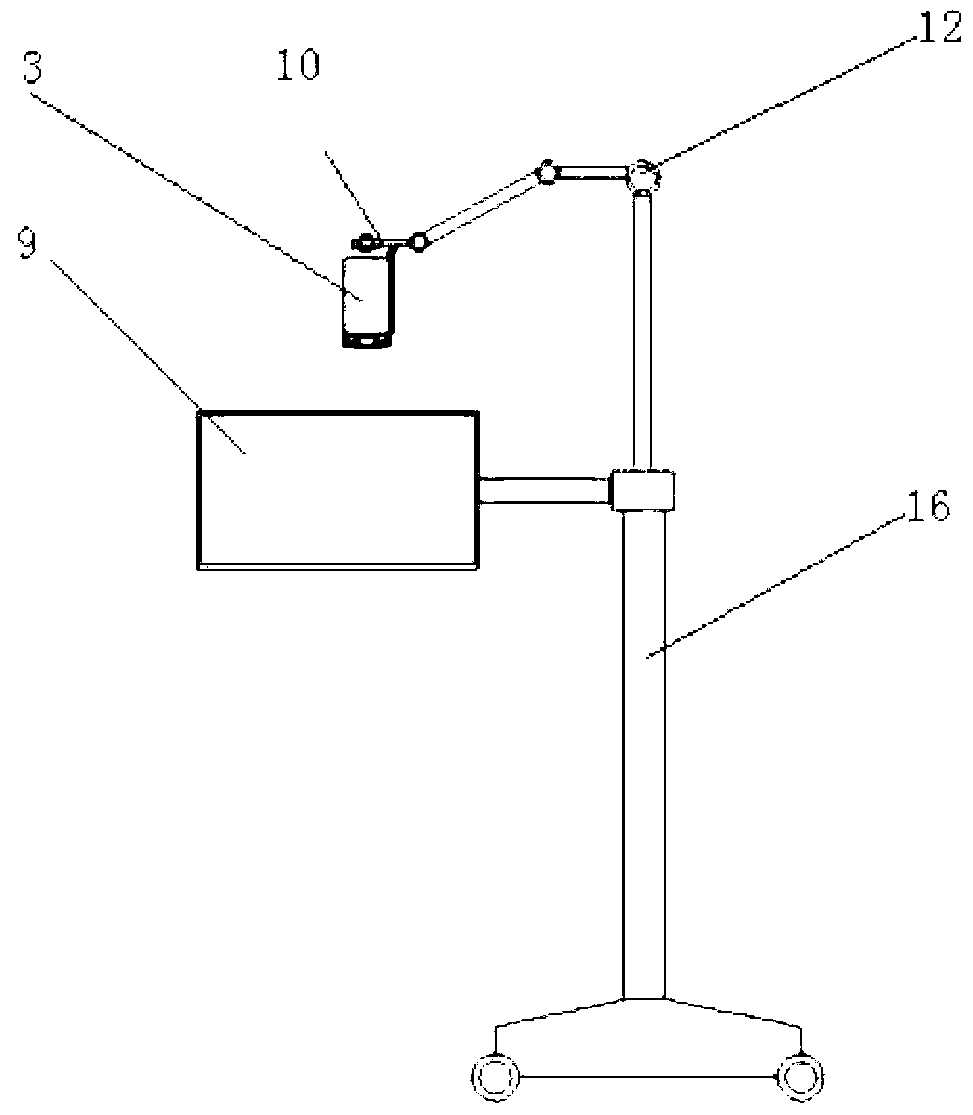
[0020] refer to figure 2 As shown, the adjustment bracket 13 of the present invention adopts a floor-type movable fixed bracket 16, and is provided with a pan-tilt...


PUM


Abstract

The invention relates to a general surgery three-dimensional micrography camera shooting presentation device which is formed by a 3D high-definition digital video, a 3D high-definition liquid crystal displayer and an adjusting support. The 3D high-definition digital video is a small integrated 3D high-definition digital video, a micrography adapter, a micrography pick-up lens and a focus light source are additionally arranged in front of an original lens of the small integrated 3D high-definition digital video, and the small integrated 3D high-definition digital video is fixed by the bed side type, suspension type or floor stand type adjusting support and connected with the 3D high-definition liquid crystal displayer. The micrography pick-up lens is composed of a single lens or a plurality of lenses, and the focal length of the micrography pick-up lens is 250mm to 500mm. The focusing light source is a cold light source which connected through light fibers or an LED light source, a focusing lens is additionally arranged at the front end of the focusing light source, and the optical axis focus of the focusing light source is identical with the focus of the 3D micrography pick-up lens. The general surgery three-dimensional micrography camera shooting presentation device is small and light, has the functions of surgery lighting, 3D real-time high-definition camera shooting, videotaping, recording, displaying, image transmitting and the like, and is suitable for multidiscipline micrography surgeries and general open surgeries in surgery systems.

Description

technical field [0001] The invention relates to a surgical operation equipment, in particular to a general surgical three-dimensional micro-camera demonstration device. Background technique [0002] Microsurgery is an important part of modern surgical techniques. With the current general-purpose surgical microscope, the operator must fix the head position for a long time, align and stare at the eyepiece at close range to perform micromanipulation, which not only easily causes operator fatigue, but also often causes cervical and lumbar spine strain. Most of the existing video recording devices for microsurgery are 2D plane images, which lack a three-dimensional effect and are not conducive to surgical teaching. Some imported surgical microscopes can be equipped with stereoscopic camera devices, such as the MCC-3000MT medical 3D high-definition camera launched by SONY at the end of 2012, which can be installed on surgical microscopes to shoot 3D images. The 3D camera is comp...

Claims


Application Information

Patent Timeline
IPC IPC(8): A61B19/00
Inventor 张湘民
Owner 上海昌宁医疗科技有限公司
Why Eureka
Try Eureka