A method for measuring multi-parameters of living mouse retina

A mouse retina and multi-parameter technology, applied in the fields of eye testing equipment, medical science, ophthalmoscope, etc., can solve the problems of slice omission, waste of funds, unfavorable animal protection, etc., and achieve accurate results, good repeatability, and easy Positioning and Quantitative Checks for Effects

Active Publication Date: 2015-09-30
EYECURE THERAPEUTICS INC JIANGSU
View PDF5 Cites 0 Cited by
  • Summary
  • Abstract
  • Description
  • Claims
  • Application Information

AI Technical Summary

Problems solved by technology

However, the eyeballs of mice are too small, and it is a difficult task that requires long-term accumulation of experience to ensure the integrity of the tissue during the slicing process; Longitudinal observational research requires the sacrifice of a large number of mice, which is a waste of funds and is not conducive to animal protection requirements

Method used


Image

  • A method for measuring multi-parameters of living mouse retina
  • A method for measuring multi-parameters of living mouse retina
  • A method for measuring multi-parameters of living mouse retina

Examples


Embodiment 1

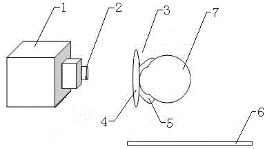
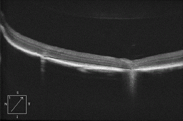
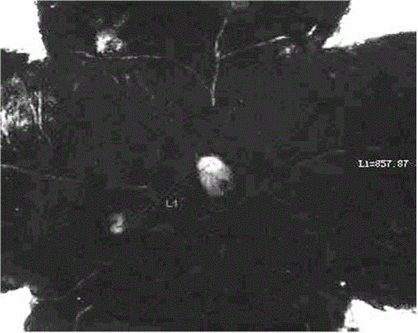
[0038] C57BL mice were selected, and attention was paid to check whether the refractive medium of the mice was transparent before the examination; at the same time, the most common clinical OCT instrument 1: Zeiss Cirrus HD-OCT 4000 or 400 was used. The anesthesia made of a mixed solution of ketamine and xylazine was injected intraperitoneally at 0.2ml / 20g body weight to anesthetize the mice. After the anesthesia was successful, laser photocoagulation was performed on the fundus of the mice, and a laser spot (diameter 50 μm) was formed at a random position on the mouse retina. , breakdown Bruch membrane). Then the mice were fixed on the three-dimensional experimental table 6; then the pupils of the mice were dilated with the mydriatic agent made of compound tropicamide diluted in normal saline, and the medical hyaluronic acid viscoelastic agent (as the concave lens 5) was covered on the On the surface of the mouse eyeball 7, take the cover glass 4 and place it on the hyaluroni...

Embodiment 2

[0043] C57BL mice were selected, and attention was paid to check whether the refractive medium of the mice was transparent before the examination; at the same time, the most common clinical OCT instrument 1: Zeiss Cirrus HD-OCT 4000 or 400 was used. Anesthesia made from a mixed solution of ketamine and xylazine was injected intraperitoneally at 0.2ml / 20g body weight to anesthetize the mice. After the anesthesia was successful, the mice were fixed on the three-dimensional experimental platform 6; After dilating the pupils of mice with a mydriatic agent made of amine, the medical hyaluronic acid viscoelastic agent (as a concave lens 5) was covered on the surface of the mouse eyeball 7, and the cover glass 4 was placed on the hyaluronic acid viscoelastic agent to form a small The plano-concave lens 3 on the front surface of the mouse eyeball 7; a Volk Superfield NC front lens 2 with a diopter of 90D is fixedly arranged in front of the lens of the OCT instrument 1, and the three-di...

Embodiment 3

[0049] C57BL mice were selected, and attention was paid to check whether the refractive medium of the mice was transparent before the examination; at the same time, the most common clinical OCT instrument 1: Zeiss Cirrus HD-OCT 4000 or 400 was used. Anesthesia made from a mixed solution of ketamine and xylazine was injected intraperitoneally at 0.2ml / 20g body weight to anesthetize the mice. After the anesthesia was successful, the mice were fixed on the three-dimensional experimental platform 6; After dilating the pupils of mice with a mydriatic agent made of amine, the medical hyaluronic acid viscoelastic agent (as a concave lens 5) was covered on the surface of the mouse eyeball 7, and the cover glass 4 was placed on the hyaluronic acid viscoelastic agent to form a small The plano-concave lens 3 on the front surface of the mouse eyeball 7; a Volk Superfield NC front lens 2 with a diopter of 90D is fixedly arranged in front of the lens of the OCT instrument 1, and the three-di...


PUM


Abstract

The invention discloses a method for measuring multiple parameters of retina of a living mouse. The method comprises the following steps: fixing a mouse, which is successfully anesthetized, on a three-dimensional experiment table; assembling a plano-concave lens, a front lens and an optical coherence tomography (OCT) instrument; adjusting the three-dimensional experiment table to ensure that the eyeballs of the mouse are positioned right ahead the front lens, and that a clear retina scanning eye fundus image appears on a display screen of the OCT instrument; selecting a high-definition one-line scanning mode to obtain a cross-sectional image of the total retina of the mouse, and measuring the thickness of each layer on the retina of the mouse; selecting a retinal volume mode, aligning the center of a cross scanning line with the optic disk of the mouse to measure the average thickness and volume of nine subareas of the retina of the mouse, and reconstructing the form of the retina of the mouse in a three-dimensional mode; and finally, selecting an optic disk scanning mode to acquire continuous thickness data of a nerve fiber layer of the retina. The method has the characteristics of high simplicity and feasibility, high repeatability, accurate result and zero external environment interference and is suitable for popularization and application.

Description

technical field [0001] The invention relates to the technical field of small rodent living retina measurement, in particular to a method for measuring multiple parameters of living mouse retina by optical coherence tomography. Background technique [0002] As an experimental animal whose genotype is highly similar to that of human, mice are widely used in medical research, as well as basic research in ophthalmology. Therefore, a mature and reliable mouse retinal measurement technique is of great significance for the study of animal models of ophthalmic diseases. At present, the measurement of the retina of mice is mainly performed by enucleating the eyes and observing histological sections after killing the mice. However, the eyeballs of mice are too small, and it is a difficult task that requires long-term accumulation of experience to ensure the integrity of the tissue during the slicing process; Longitudinal observational studies require the sacrifice of a large number ...

Claims


Application Information

Patent Timeline
Patent Type & Authority Patents(China)
IPC IPC(8): A61B3/14A61B3/12
Inventor 袁松涛宋清露计江东孙杏红聂桥
Owner EYECURE THERAPEUTICS INC JIANGSU
Why Eureka
Try Eureka