Composite multifunctional biological hemostatic material and preparation method thereof
A technology of hemostatic materials and biomaterials, which is applied in the field of medical devices and can solve the problems of no hemostatic materials
- Summary
- Abstract
- Description
- Claims
- Application Information
AI Technical Summary
Problems solved by technology
Method used
Image
Examples
Embodiment 1
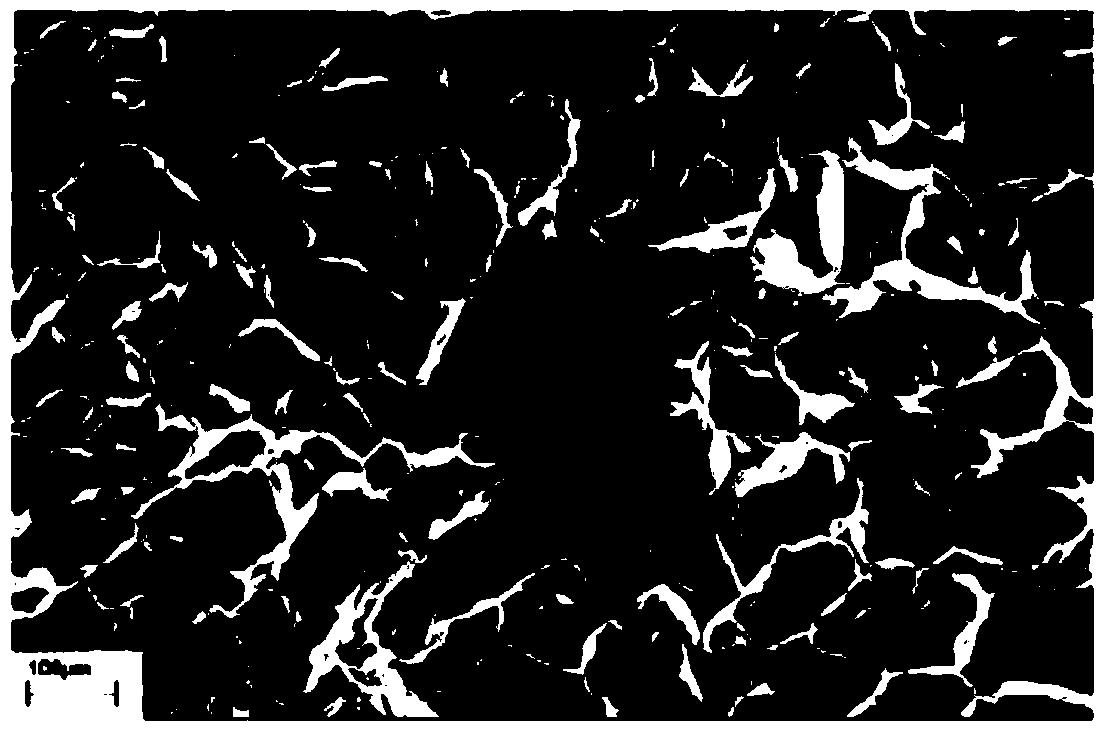
[0034] (1) Prepare 5wt% silk fibroin aqueous solution with a molecular weight of 5000, pour it into the mold, adjust the freezing rate of the freeze dryer to 0.1°C / min, freeze-drying temperature -45°C, freeze-drying and drying time for 2 hours, sublimation The temperature is 10°C, and finally the temperature is raised to 30-40°C until the pressure is 0.5 Pa / min, and the freeze-drying process is completed to obtain a silk fibroin scaffold (that is, a porous tissue cell growth scaffold). The scanning electron microscope picture is shown in figure 1 .
[0035](2) Prepare 4wt% chitosan aqueous solution with a molecular weight of 10,000, soak the silk fibroin support prepared in the previous step in the chitosan aqueous solution, place them in the mold together, and adjust the freezing rate of the freeze dryer at 2°C / The freeze-drying temperature is -15°C, the freeze-drying drying time is 3 hours, the sublimation temperature is 20°C, and finally the temperature is raised to 30-40°...
Embodiment 2
[0037] (1) Prepare 20wt% silk fibroin aqueous solution with a molecular weight of 8000, pour it into the mold, adjust the freezing rate of the freeze dryer to 1°C / min, freeze-drying temperature -30°C, freeze-drying and drying time for 4 hours, sublimation The temperature is 15°C, and finally the temperature is raised to 30-40°C until the pressure is 2Pa / min, and the freeze-drying process is completed to obtain a silk fibroin scaffold (that is, a porous tissue cell growth scaffold). The scanning electron microscope picture is shown in image 3 .
[0038] (2) Prepare 30wt% chitosan aqueous solution with a molecular weight of 30,000, soak the silk fibroin support prepared in the previous step in the chitosan aqueous solution, place them in the mold together, and adjust the freezing rate of the freeze dryer to 1.5°C / The freeze-drying temperature is -30°C, the freeze-drying drying time is 2.5 hours, the sublimation temperature is 10°C, and finally the temperature is raised to 30-4...
Embodiment 3
[0040] 1) Prepare 20wt% silk fibroin aqueous solution with a molecular weight of 8000, pour it into the mold, adjust the freezing rate of the freeze dryer to 1°C / min, the freeze-drying temperature to -30°C, the freeze-drying time to 4 hours, and the sublimation temperature 15°C, and finally raise the temperature to 30-40°C until the pressure is 2Pa / min, complete the freeze-drying process, and obtain the silk fibroin scaffold (that is, the porous tissue cell growth scaffold).
[0041] 2) prepare 30wt% molecular weight chitosan aqueous solution of 30,000, which contains 5wt% PVP, soak the silk fibroin support prepared in the previous step in the chitosan aqueous solution, place them in the mold together, and adjust the freeze dryer The freezing rate is 1.5°C / min, the freeze-drying temperature is -30°C, the freeze-drying drying time is 2.5 hours, the sublimation temperature is 10°C, and finally the temperature is raised to 30-40°C until the pressure is 2Pa / min. Silk fibroin-chito...
PUM
| Property | Measurement | Unit |
|---|---|---|
| pore size | aaaaa | aaaaa |
Abstract
Description
Claims
Application Information
 - R&D
- Intellectual Property
- Life Sciences
- Materials
- Tech Scout
- Unparalleled Data Quality
- Higher Quality Content
- 60% Fewer Hallucinations
Browse by: Latest US Patents, China's latest patents, Technical Efficacy Thesaurus, Application Domain, Technology Topic, Popular Technical Reports.
© 2025 PatSnap. All rights reserved.Legal|Privacy policy|Modern Slavery Act Transparency Statement|Sitemap|About US| Contact US: help@patsnap.com


