Method for marking immune globulin by quantum dot

A technology of immunoglobulin and quantum dots, which is applied in the field of quantum dot-labeled immunoglobulins, can solve the problems of large loss of immunoglobulins, reduced activity, and low efficiency, and achieves simple operation and use of equipment, improved utilization, and high activity. high effect

Active Publication Date: 2014-01-01
SHENZHEN KINGFOCUS BIOMDICAL ENG CO LTD
View PDF10 Cites 5 Cited by
  • Summary
  • Abstract
  • Description
  • Claims
  • Application Information

AI Technical Summary

Problems solved by technology

[0004] The technical problem solved by the present invention is to provide a method for amplifying industrial production of quantum dot-labeled immunoglobulin, and solve the problems of greatly reduced activity of immunoglobulin after labeling, large loss of immunoglobulin, low efficiency and high cost

Method used


Image

  • Method for marking immune globulin by quantum dot
  • Method for marking immune globulin by quantum dot
  • Method for marking immune globulin by quantum dot

Examples


Embodiment 1

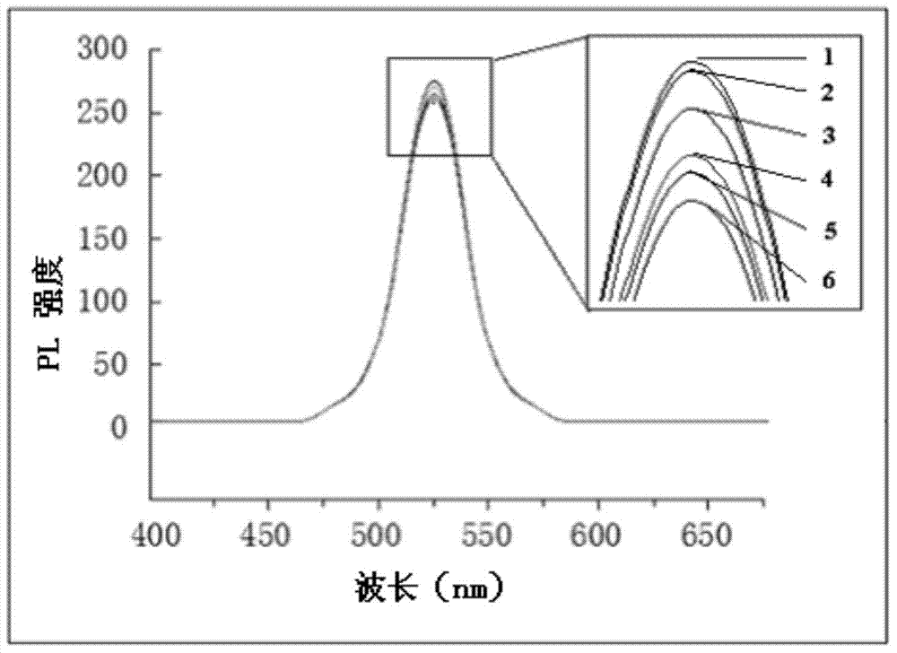
[0026] A CdTe / ZnS core-shell quantum dot-labeled Procalcitonin (PCT) antibody with a fluorescence wavelength of 525 nm was selected.

[0027] (1) Cleaning quantum dots: Take 10 μL of 8 μmol / mL CdTe / ZnS core-shell quantum dots in EP (eppendorf) tubes, and add 2 mL of 10 mmol / L, pH=7.2 phosphate buffer into the EP tubes, mix well and use The 100K ultrafiltration tube was centrifuged at 5000r / min at 25°C for 6min, and the quantum dots were recovered in EP tubes. Repeat the above steps three times, and make the total volume of the quantum dot solution not exceed 50 μL for the last time. Use a micropH meter to detect the pH of the quantum dot solution and adjust the pH value to 7.2.

[0028] (2) Activation: Weigh 2 mmol of EDC and 0.5 mmol of NHS in an ice box with humidity less than or equal to 30% and 0-4°C, and quickly add them to the quantum dot solution obtained in step (1), and vortex After oscillating evenly, place on a shaker to react for 50 minutes at a reaction temperat...

Embodiment 2

[0036] (1) Cleaning of quantum dots: Take 10 μL of 8 μmol / mL CdTe / ZnS core-shell quantum dots in EP tube, and add 2 mL of 10 mmol / L, pH=7.5 phosphate buffer into the EP tube, mix well and use 100K ultrafiltration The tube was centrifuged at 8000r / min at 25°C for 8min, and the quantum dots were recovered in EP tubes. Repeat the above steps three times, and make the total volume of the quantum dot solution not exceed 50 μL for the last time. Use a micropH meter to detect the pH of the quantum dot solution and adjust the pH value to 7.5.

[0037] (2) Activation: Weigh 2.5mmol EDC and 0.5mmol NHS in an ice box with a humidity less than or equal to 30% and 0-4°C and quickly add them to the quantum dot solution obtained in step (1), shake evenly with a vortex oscillator Then place it on a shaker and react for 80 minutes at a reaction temperature of 25° C. to obtain an activated quantum dot solution.

[0038] (3) Second cleaning of quantum dots: Add 2mL of 50mmol / L, pH=8.5 borate b...
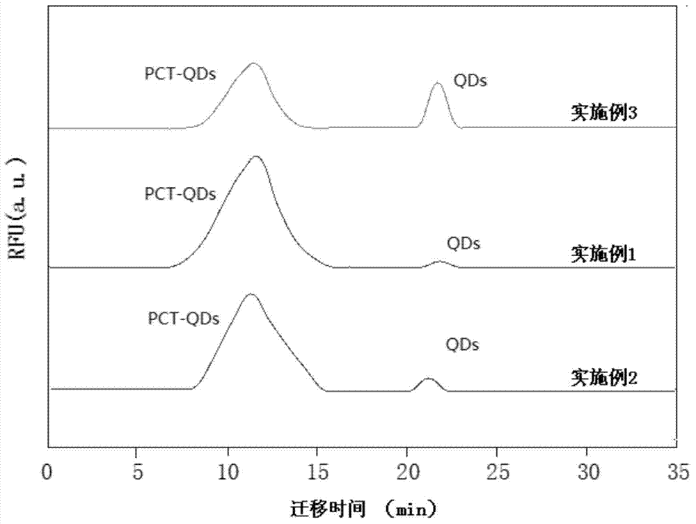
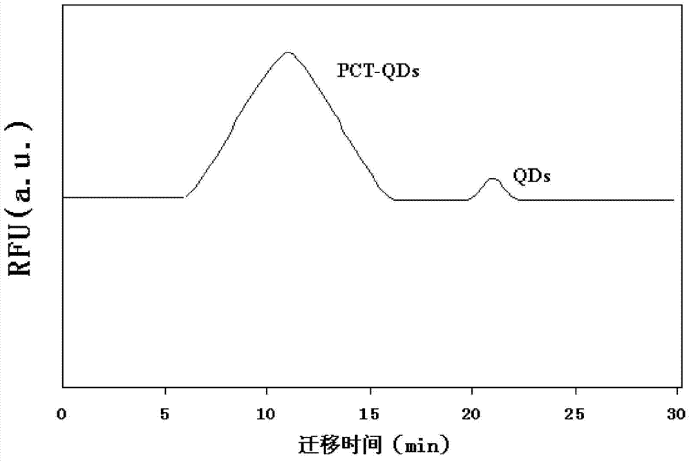
Embodiment 3

[0044] The difference between this implementation and Examples 1 and 2 is that the labeling efficiency is significantly reduced by using labeling conditions outside the range of the key parameters in the specification.

[0045] (1) Cleaning quantum dots: Take 10 μL, 8 μmol / mL CdTe / ZnS core-shell quantum dots in the EP tube, and add 2 mL, 10 mmol / L, pH=7.4 phosphate buffer into the EP tube, mix well Use a 100K ultrafiltration tube to centrifuge at 7000 r / min at 25°C for 8 min, and recover the quantum dots in EP tubes. Repeat the above steps three times, and make the total volume of the quantum dot solution not exceed 50 μL for the last time. Use a micropH meter to detect the pH of the quantum dot solution and adjust the pH value to 7.5.

[0046] (2) Activation: Weigh 2.5mmol EDC and 0.5mmol NHS in an ice box with a humidity less than or equal to 30% and 0-4°C and quickly add them to the quantum dot solution obtained in step (1), shake evenly with a vortex oscillator Then plac...


PUM


Abstract

The invention belongs to the field of a nano-biotechnology and particularly relates to a method for marking immune globulin by a quantum dot. The method comprises the following steps: washing the quantum dot, activating, washing the quantum dot for the second time, washing the immune globulin, coupling and separating. According to the method, structural characteristics of the water-soluble quantum dot, the reaction characteristics of EDC (1-(3-Dimethylaminopropyl)-3-ethylcarbodiimide hydrochloride) activated carboxyl and the reaction characteristics of a condensation reaction are utilized; the carboxyl on the surface of the quantum dot is activated sufficiently by optimizing conditions of all the steps and is in a suitable marking environment; the quantum dot can be combined with the surface of the immune globulin by the condensation reaction through optimizing the condition of the coupling step, and the marking efficiency is high. The immune globulin which is not reacted can be recycled to be marked continually through exclusion chromatography, separation and purification by a reaction solution obtained by the coupling step so that the utilization rate of the immune globulin is improved and the cost is saved. According to the method provided by the invention, the activity of a prepared protein-quantum dot conjugate is high, the operation and used equipment are simple, and the production can be enlarged industrially.

Description

technical field [0001] The invention relates to the field of nanobiology technology, in particular to a method for marking immunoglobulin with quantum dots. Background technique [0002] Quantum dots (QDs) are aggregates composed of a certain number of actual atoms, and semiconductor compounds with three-dimensional dimensions less than 100nm. Quantum dots have many advantages such as high luminous intensity, wide excitation spectrum, narrow emission spectrum, long fluorescence lifetime, multifunctional surface modification and good stability. They have the potential to replace traditional organic fluorescent dyes in the field of fluorescence detection and have become a new generation of biological Fluorescent markers. In various immunological experiments, the method of tracking proteins and detecting protein content by labeling fluorescent molecules on proteins has been widely used. [0003] In 2009, Zhang Guohua et al. published a method for labeling ractopamine antibodi...

Claims


Application Information

Patent Timeline
IPC IPC(8): G01N33/533
CPCG01N33/533A61K49/0067C07K16/00G01N33/588G01N33/6854
Inventor 张二盈章国建
Owner SHENZHEN KINGFOCUS BIOMDICAL ENG CO LTD
Why Eureka
Try Eureka