Method for determining free analyte in biological sample and determining drug protein binding ratio

A technology for biological samples and analytes, which is applied in the field of chemical analysis, can solve problems such as limitations and limitations on the application of pre-equilibrium kinetic correction methods, and achieve the effects of low cost, short extraction time, and simple equipment

Inactive Publication Date: 2015-05-27
SHENZHEN POLYTECHNIC
View PDF0 Cites 0 Cited by
  • Summary
  • Abstract
  • Description
  • Claims
  • Application Information

AI Technical Summary

Problems solved by technology

These factors limit the application of the pre-equilibrium kinetic correction method in microextraction technology, and also limit the application of the pre-equilibrium kinetic correction hollow fiber liquid phase microextraction method in the rapid analysis of free drugs

Method used


Image

  • Method for determining free analyte in biological sample and determining drug protein binding ratio
  • Method for determining free analyte in biological sample and determining drug protein binding ratio
  • Method for determining free analyte in biological sample and determining drug protein binding ratio

Examples


preparation example Construction

[0066] In the preparation of the solvent rod in step b, one end of the hollow fiber membrane is heated and sealed for 2 mm with tweezers, and then 50 μL of extraction solvent (such as n-octanol) is slowly injected into the hollow fiber membrane with a 50 μL flat-tip micro-syringe. Set aside for 10 min, then cut off the hollow fiber membrane 2 mm above the liquid surface, and then heat and seal the open end with tweezers for about 2 mm.

[0067] Step 2, through the back extraction process of different concentrations of standard analytes, obtain the time constant of the solvent rod hollow fiber liquid phase micro-extraction by the concentration curve method: place the solvent rod prepared in step 1 in the 1-5 mL sample that has been loaded. solution vials with the solvent stick fully immersed in the sample matrix: for example, the vial can be 2 mL in size and the sample solution is 1.8 mL, so that the solvent stick is fully immersed in the sample solution; place the vial in On t...

Embodiment 1

[0087] Cut polyvinylidene fluoride (PVDF) hollow fiber membranes (pore size 0.2 μm, inner diameter 1.2 mm, outer diameter 1.4 mm) into small sections of 30 mm, and ultrasonically clean them with methanol for 2 min to remove impurities on the surface of the hollow fiber membranes. Air-dry the hollow fiber membrane in a cabinet and set aside.

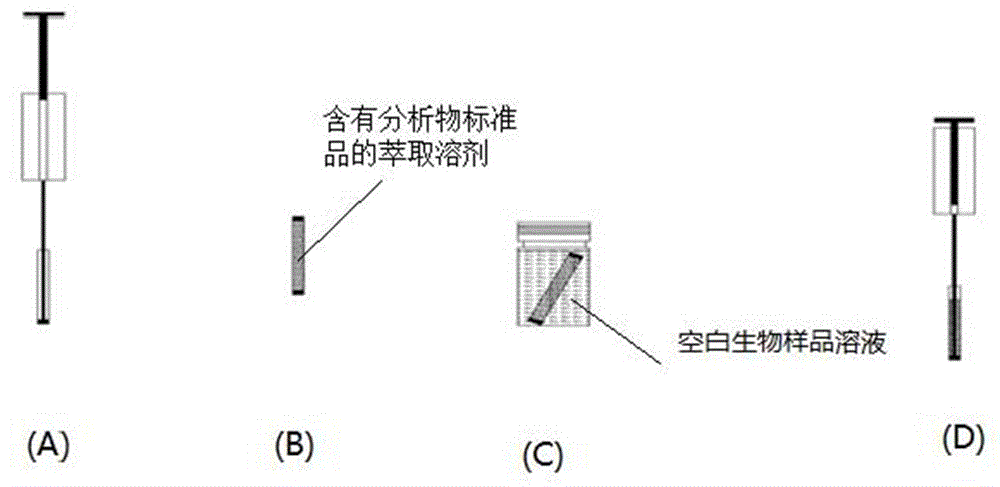
[0088]Concentration curve determination of the time constant in phosphate buffer solution: One end of the prepared hollow fiber membrane was heated and sealed for 2 mm with tweezers, and then slowly injected into the hollow fiber membrane with a 50 μL flat-tip microsyringe, 50 μL containing different concentrations of Ve. Local anesthetics (0.5, 1, 2, 3, 4, 5 μg / mL lidocaine hydrochloride, bupivacaine hydrochloride, and tetracaine hydrochloride) in n-octanol extract solutions (eg figure 1 A), stand for 10 min, the extraction solution fills the wall pores of the hollow fiber membrane, and the hollow fiber membrane is cut off 2 mm above the...

Embodiment 2

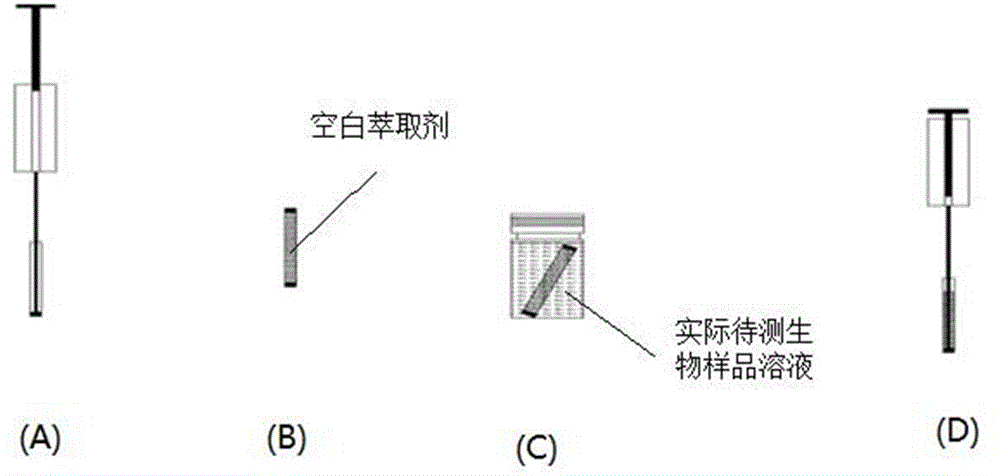
[0099] The preparation and operation process of the solvent rod hollow fiber liquid phase microextraction device are the same as those in Example 1. First, solvent bars containing 10, 20, 25, 30, 40, and 50 μg / mL of local anesthetics (lidocaine hydrochloride, bupivacaine hydrochloride, and tetracaine hydrochloride) in n-octanol extraction solutions were placed in blank 1.8 mL blank bovine serum albumin in phosphate buffer solution (wherein the concentration of bovine serum albumin in the phosphate buffer solution is 1% (w / v)) for 12 min, draw a Q-q0 back-extraction concentration curve to obtain a Linear equation, using the slope of the linear equation, to find the time constant a according to equation (1); Extract the albumin in phosphate buffer solution for 12 min to obtain the extraction amount n of the local anesthetic; finally, substitute the time constant a and the extraction amount n into formula (2) to obtain the free concentration Cf of the local anesthetic in the bovi...


PUM

PropertyMeasurementUnit
pore sizeaaaaaaaaaa

Abstract

The invention discloses a method for determining a free analyte in a biological sample. The method comprises the following steps: adopting the solvent bar hollow fiber liquid-phase micro-extraction technology, determining a time constant of the solvent bar hollow fiber liquid-phase micro-extraction through a concentration curve method in the reverse extraction process, and further computing the concentration of the free analyte in the actual sample through the analyte pre-equilibrium concentration obtained through determining the pre-equilibrium solvent bar hollow fiber liquid-phase micro-extraction. In the biological sample containing protein, the drug protein binding ratio can be computed according to the free drug concentration. According to the method, the sample pretreatment device is simple and convenient to operate. The deuterated material is not required, the property of the biological sample is not changed, the content of the free analyte in the biological sample can be determined simply, quickly and accurately.

Description

technical field [0001] The invention relates to a chemical analysis method, in particular to a method for measuring free analytes in biological samples and a method for measuring the binding rate of drug proteins. Background technique [0002] In recent years, free drugs have received more and more attention and attention, and have become one of the research hotspots in pharmacokinetics and therapeutic drug monitoring. After the drug enters the bloodstream of the body, it exists in two forms. One is a conjugated drug; weakly acidic drugs bind to albumin, and weakly basic drugs bind to 1-acid glycoprotein (AGP). The other is the free drug. The free drug in plasma is the part that produces pharmacological effects. The concentration of free drug is in balance with the drug at the receptor site, so it is closely related to drug efficacy or adverse drug reactions. Only free drugs can be transformed and excreted by the body. [0003] Hollow fiber liquid phase microextraction pl...

Claims


Application Information

Patent Timeline
Patent Type & Authority Patents(China)
IPC IPC(8): G01N30/00G01N30/08
Inventor 崔淑芬张英雪陈敏侯金星
Owner SHENZHEN POLYTECHNIC
Why Eureka
Try Eureka