

Double-labeled immunohistochemical staining kit for micro invasive lung adenocarcinoma
A technique for immunohistochemistry and staining reagents, which is used in measuring devices, instruments, disease diagnosis, etc., to achieve the effects of short experiment time, fewer staining steps, and intuitive comparison of experimental results.
- Summary
- Abstract
- Description
- Claims
- Application Information
AI Technical Summary
Problems solved by technology
Method used
Image

Examples
Embodiment 1
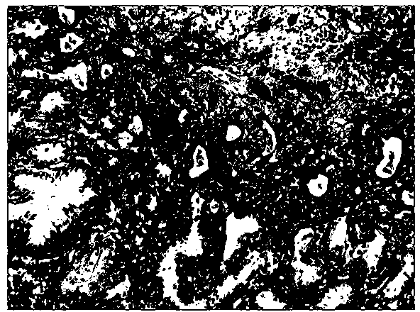
[0027] The research objects were formaldehyde-fixed-paraffin-embedded microinvasive lung adenocarcinoma tissues (Department of Pathology, Fuzhou General Hospital of Nanjing Military Region). The steps of immunohistochemical experiment are as follows:
[0028] (1) The tissue slices were dry-baked in a 67°C incubator for 2 hours.
[0029] (2) Conventional xylene dewaxing 3 times, 6 minutes each time, hydration in 100%, 100%, 95%, 85% gradient ethanol, 3 minutes each time, and finally rinsed with tap water.
[0030] (3) High-pressure repair in citric acid antigen retrieval solution of pH 6.0 for 1.5-2 minutes, naturally cool to room temperature, rinse with tap water, and rinse with PBS for 3×3 minutes.
[0031] (4) Incubate in 3% hydrogen peroxide at room temperature for 10 minutes to block endogenous peroxidase, wash with PBS for 3×3 minutes.
[0032] (5) Block with normal animal serum for 10 minutes, shake off excess serum without washing, add CD34 and β-Tubulin-Ⅲ mixed prima...
Embodiment 2
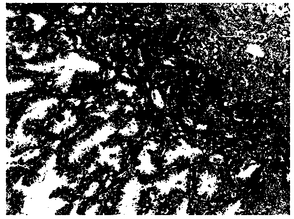
[0039] The research objects were formaldehyde-fixed-paraffin-embedded microinvasive lung adenocarcinoma tissues (Department of Pathology, Fuzhou General Hospital of Nanjing Military Region). The steps of immunohistochemical experiment are as follows:
[0040] (1) The tissue slices were dry-baked in a 67°C incubator for 2 hours.
[0041] (2) Conventional xylene dewaxing 3 times, 6 minutes each time, hydration in 100%, 100%, 95%, 85% gradient ethanol, 3 minutes each time, and finally rinsed with tap water.
[0042] (3) High-pressure repair in citric acid antigen retrieval solution of pH 6.0 for 1.5-2 minutes, naturally cool to room temperature, rinse with tap water, and rinse with PBS for 3×3 minutes.
[0043] (4) Incubate in 3% hydrogen peroxide at room temperature for 10 minutes to block endogenous peroxidase, wash with PBS for 3×3 minutes.
[0044] (5) Block with normal animal serum for 10 minutes, shake off excess serum without washing, add CD34 and β-Tubulin-Ⅲ mixed prima...
Embodiment 3
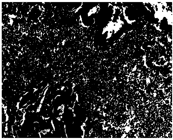
[0051] The research objects were formaldehyde-fixed-paraffin-embedded microinvasive lung adenocarcinoma tissues (Department of Pathology, Fuzhou General Hospital of Nanjing Military Region). The steps of immunohistochemical experiment are as follows:
[0052] (1) The tissue slices were dry-baked in a 67°C incubator for 2 hours.
[0053] (2) Conventional xylene dewaxing 3 times, 6 minutes each time, hydration in 100%, 100%, 95%, 85% gradient ethanol, 3 minutes each time, and finally rinsed with tap water.
[0054] (3) High-pressure repair in citric acid antigen retrieval solution of pH 6.0 for 1.5-2 minutes, naturally cool to room temperature, rinse with tap water, and rinse with PBS for 3×3 minutes.
[0055] (4) Incubate in 3% hydrogen peroxide at room temperature for 10 minutes to block endogenous peroxidase, wash with PBS for 3×3 minutes.
[0056] (5) Block with normal animal serum for 10 minutes, shake off excess serum without washing, add CD34 and β-Tubulin-Ⅲ mixed prima...
PUM
 Abstract
Description
Claims
Application Information