CT liver-perfusion image post-processing method and CT liver-perfusion method

A technology of liver perfusion and post-processing, which is applied in the field of medical devices and can solve problems such as the time curve of wrong contrast agent concentration, the influence of calculation results of perfusion parameters, and large changes in the peak value of image gray values.

Active Publication Date: 2014-06-04
SHANGHAI UNITED IMAGING HEALTHCARE
View PDF5 Cites 16 Cited by
  • Summary
  • Abstract
  • Description
  • Claims
  • Application Information

AI Technical Summary

Problems solved by technology

However, for different patients and different contrast agent injection doses and speeds, the peak value of the image gray value of the blood vessel area varies greatly. When operating manually, it is ea

Method used


Image

  • CT liver-perfusion image post-processing method and CT liver-perfusion method
  • CT liver-perfusion image post-processing method and CT liver-perfusion method
  • CT liver-perfusion image post-processing method and CT liver-perfusion method

Examples


no. 1 example

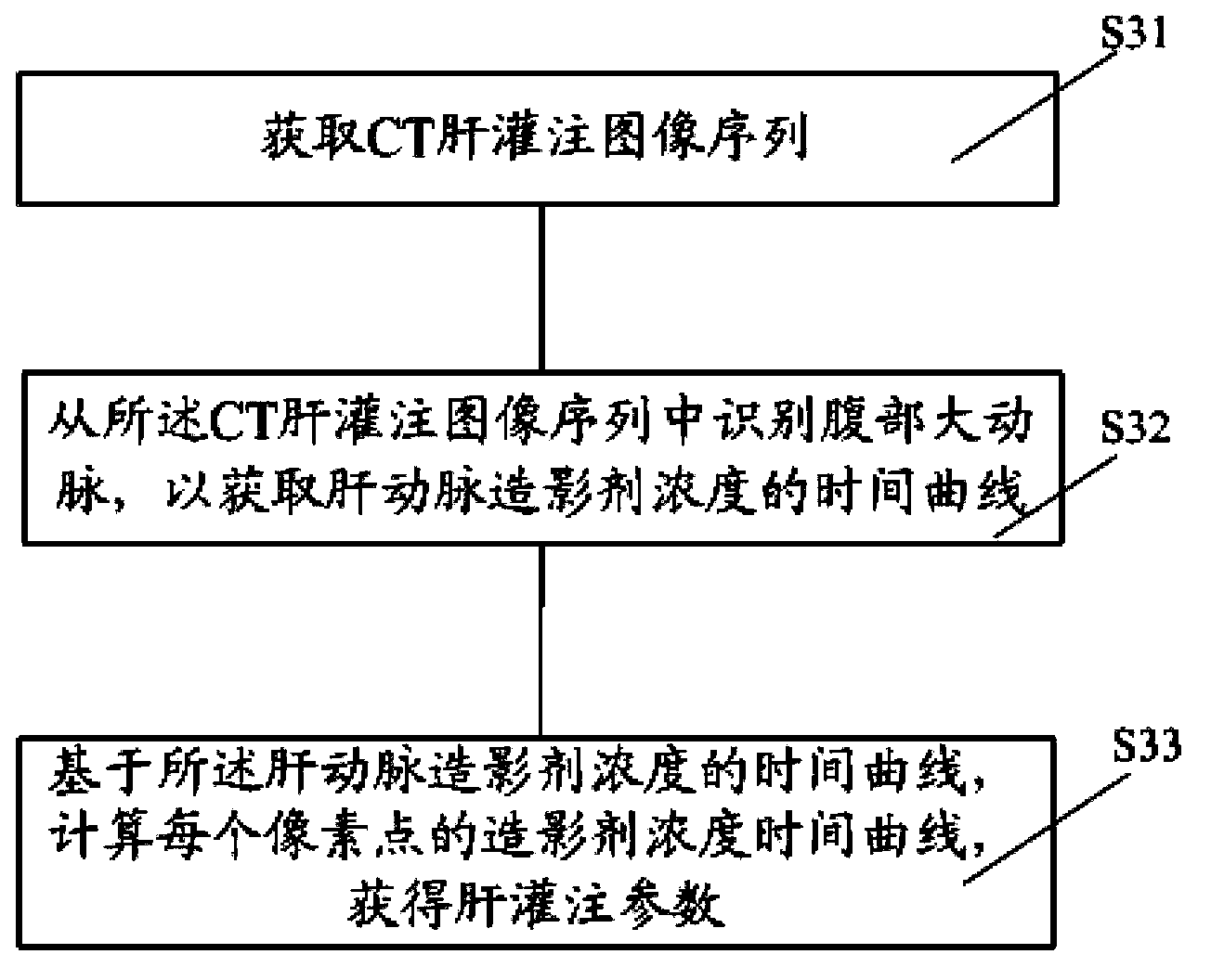
[0040] The first embodiment of the image post-processing method of CT liver perfusion provided by the present invention includes:

[0041] Step S31 is executed to obtain a sequence of CT liver perfusion images. The present invention forms a CT liver perfusion image sequence by way of CT scanning. In this embodiment, a CT liver perfusion image sequence is acquired from a CT scanner. Since a CT scan of the liver is usually performed every 1-4 seconds, a total of 15-20 scans are performed, therefore, 15-20 groups of three-dimensional volume data about the human liver can be obtained from the CT scanner, and the 15 ~20 groups of three-dimensional volume data correspond to the information collected by CT scans on the human liver at different time points, and are arranged according to time to obtain CT liver perfusion image sequences.

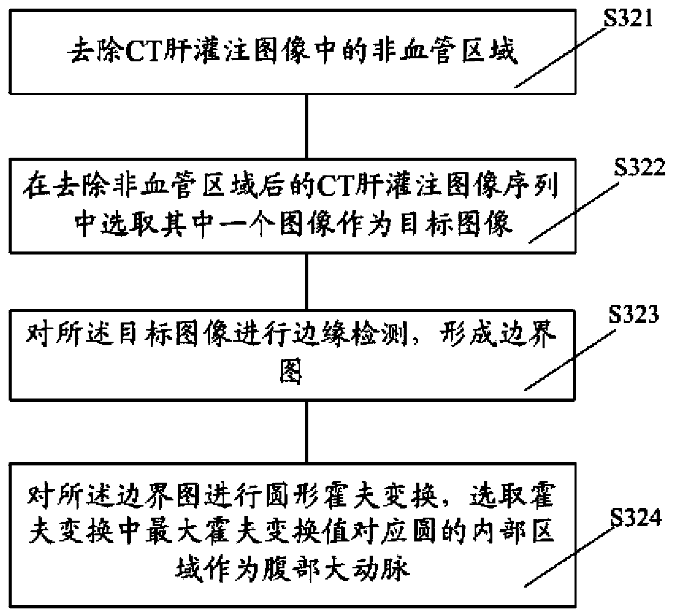
[0042] Step S32 is executed to identify the abdominal aorta from the CT liver perfusion image sequence, so as to obtain the time curve of contrast...


PUM


Abstract

The invention provides a CT liver-perfusion image post-processing method and a CT liver-perfusion method. The CT liver-perfusion image post-processing method includes: obtaining a CT liver-perfusion image sequence and identifying an abdominal aorta from the CT liver-perfusion image sequence so as to obtain a time curve of a hepatic-artery contrast-agent concentration; and based on the time curve of the hepatic-artery contrast-agent concentration, calculating a contrast-agent concentration time curve of each pixel point and obtaining liver-perfusion parameters. The invention also provides a CT liver-perfusion method which uses the CT liver-perfusion image post-processing method. The CT liver-perfusion image post-processing method and the CT liver-perfusion method are higher in accuracy.

Description

technical field [0001] The invention relates to the field of medical devices, in particular to an image post-processing method of CT liver perfusion and a CT liver perfusion method. Background technique [0002] Perfusion refers to the function of blood to transport oxygen and other substances carried by blood to surrounding tissues through the network of capillaries. Hepatic perfusion refers to the process in which blood passes through the hepatic artery, hepatic portal vein and hepatic sinusoids, and the oxygen and other substances in it are transported to the liver tissue for utilization or transformation, which is generally equivalent to the blood flow process. [0003] Electronic computer X-ray tomography (CT) perfusion imaging is to scan the arterial layer of the selected layer after intravenous injection of contrast agent to obtain the time-density curve of each pixel in the layer ( time-density curve, TDC), and then according to the curve, calculate perfusion parame...

Claims


Application Information

Patent Timeline
IPC IPC(8): G06T7/00G06F19/00
CPCG06T7/0012G06T2207/10016G06T2207/10081G06T2207/20061G06T2207/30056
Inventor 王潚崧
Owner SHANGHAI UNITED IMAGING HEALTHCARE
Why Eureka
Try Eureka