
Application of microRNA in serum serving as diagnostic marker of liver cancer

A technology for diagnosing markers and liver cancer, applied in the fields of biotechnology and medicine, can solve the problems of small sample size, lack of early-stage liver cancer diagnostic markers, lack of verification, etc., and achieve good sensitivity and specificity.

Active Publication Date: 2014-07-16
青岛瑞思德医学检验实验室有限公司
View PDF1 Cites 4 Cited by
  • Summary
  • Abstract
  • Description
  • Claims
  • Application Information

AI Technical Summary

Problems solved by technology

The above results suggest that the diagnosis of HCC by serum microRNA is feasible, but there are many defects in these studies, such as the methods used to screen microRNA and the number of candidate genes are limited, the diagnosis of liver cancer at different stages is not detected, the sample size is small, and there is no early liver cancer. diagnostic markers or lack of independent validation

Method used


Image

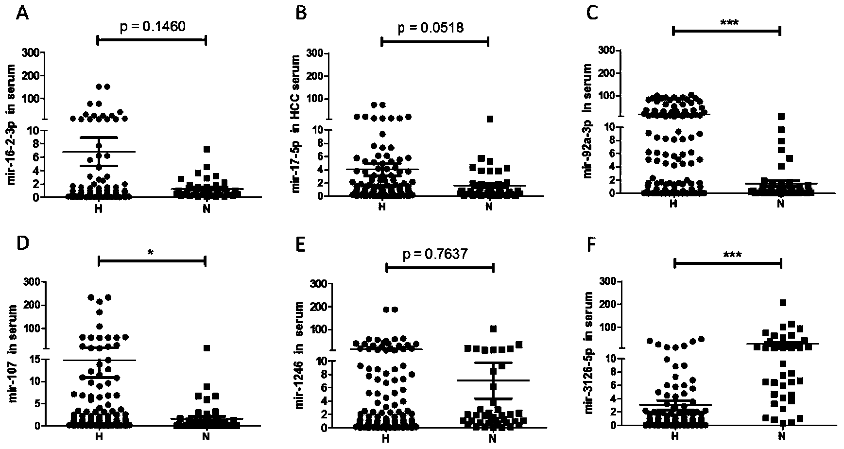
  • Application of microRNA in serum serving as diagnostic marker of liver cancer
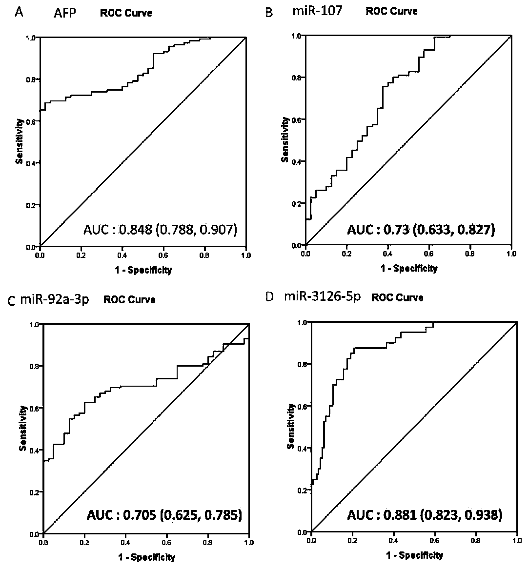
  • Application of microRNA in serum serving as diagnostic marker of liver cancer
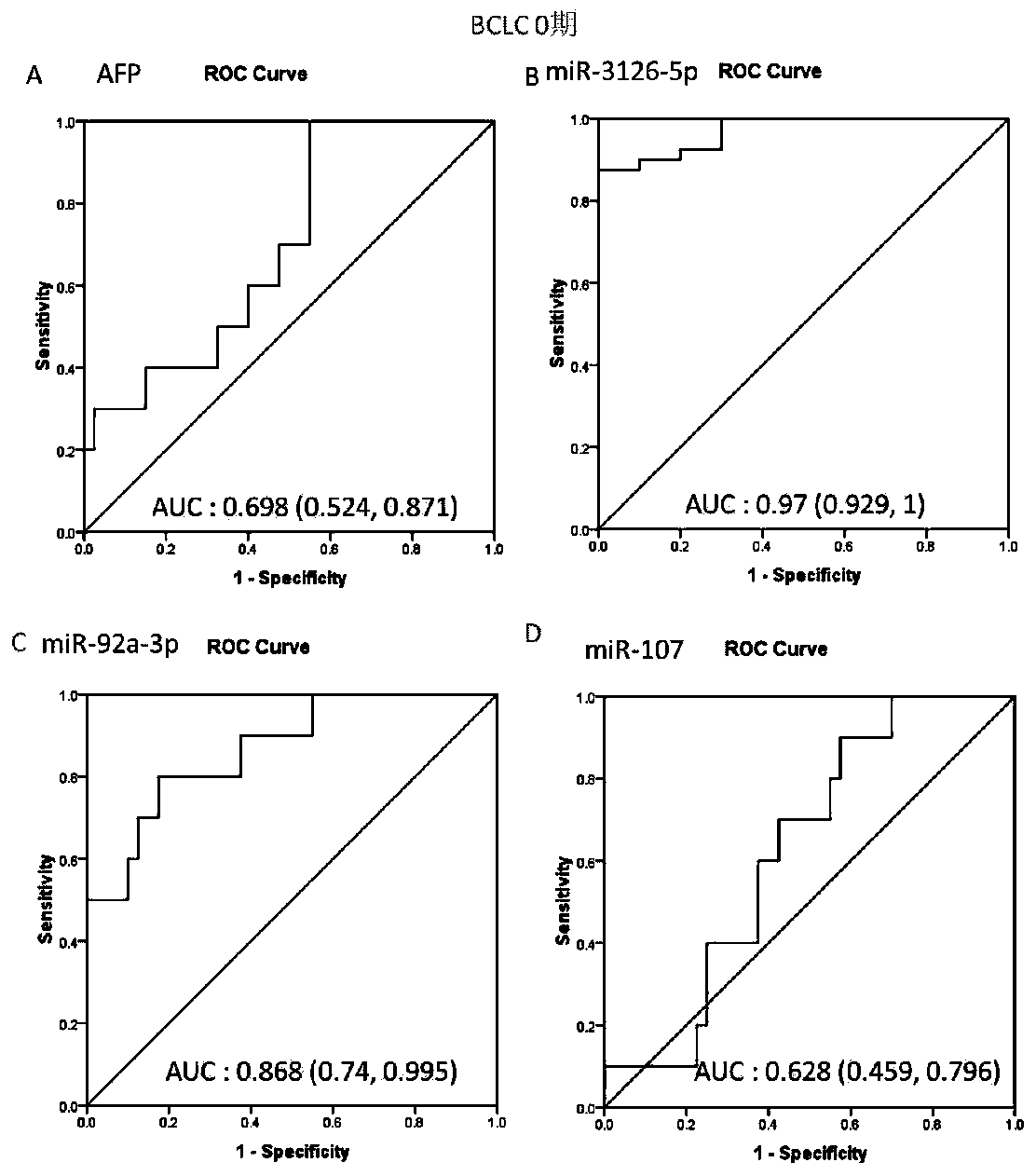
  • Application of microRNA in serum serving as diagnostic marker of liver cancer

Examples


Embodiment 1

[0044] Example 1 Serum Sample Collection

[0045] A total of 155 serum samples were collected, including 50 normal human serum and 105 liver cancer patient serum. Serum was collected from the Department of Infectious Diseases, Provincial Hospital of Shandong University. Serum was collected at the time of clinical diagnosis, from March 2012 to November 2013. Serum was collected in coagulation-promoting tubes, centrifuged at 2000-3000rpm for 18-20min, the supernatant was drawn into centrifuge tubes, and stored at -80°C. The clinical case characteristics of the patients are shown in Table 2.

[0046] Table 2 Clinical liver cancer cases and their clinical features

[0047]

[0048] Tumor staging was based on the Barcelona grading standard, 0 and A in BCLC classification were early stage, and B, C and D in BCLC classification were advanced stage.

Embodiment 2

[0049] Example 2 Extraction of serum microRNA

[0050] Proceed as follows:

[0051] (1) Aspirate 400 μl of frozen patient serum, add 750 μl of lysate MRL, and pipette several times.

[0052] (2) Shake the homogenate sample vigorously, incubate at room temperature for 5 min, and carefully transfer the supernatant to a new RNase-free centrifuge tube.

[0053] (3) Add 200 μl chloroform for every 750 μl lysate, cover the sample tube tightly, shake vigorously for 15 seconds and incubate at room temperature for 3 minutes.

[0054] (4) Centrifuge at 12000rpm at 4°C for 10 minutes; the sample will be divided into 3 layers, remove the upper aqueous phase, and transfer to a new RNase free EP tube.

[0055] (5) Add 0.6 times the volume of 70% ethanol, invert and mix well, and transfer the resulting solution and possible precipitates into the adsorption column RA.

[0056] (6) Centrifuge at 10000rpm for 45s, collect the filtrate, add 2 / 3 volume of absolute ethanol, mix by inversion, p...

Embodiment 3

[0061] Example 3 microRNA reverse transcription

[0062] Take 2 μg RNA, thaw at room temperature, and put it on ice immediately after thawing. Prepare the mixture according to Table 3.

[0063] Table 3 Reaction system for microRNA reverse transcription

[0064]

[0065] 37°C, 60min; 85°C, 5min, then put on ice, the obtained microRNA cDNA can be used for subsequent experiments, or stored at -20°C.


PUM


Abstract

The invention discloses an application of microRNA-miR-3126-5p in serum serving as a diagnostic marker of liver cancer. In the invention, a high-throughput chip is used for detecting expression of microRNA in liver cancer cells at different development stages and in clinical liver cancer serum samples, candidate microRNA is selected, the expression of the candidate microRNA in the serum samples is detected to find that the miR-3126-5p has good sensitivity and specificity on diagnosis of liver cancer and especially has a good diagnostic value in diagnosis of early liver cancer and liver cancer with low AFP expression. On the basis, the invention develops a primer, a kit and a detection method for detecting the miR-3126-5p, which have good development prospects and clinical application values. The sequence of the primer is as shown in SEQ ID NO.2, and the kit is composed of a microRNA extraction reagent, a reverse transcription reagent, a specific amplification primer and a PCR amplification reagent.

Description

technical field [0001] The invention relates to the application of microRNA in serum—miR-3126-5p as a liver cancer diagnostic marker, and belongs to the fields of biotechnology and medicine. Background technique [0002] Primary hepatocellular carcinoma (hepatocellular carcinoma, HCC) is one of the most common malignant tumors in my country. According to relevant statistics, the annual new cases and death rate of male liver cancer patients rank among the top five, and the death rate of female liver cancer patients ranks seventh. Hepatocellular carcinoma is the main pathological type of liver cancer, accounting for about 80% of primary liver cancer and 90% in our country. One of the main reasons for its high mortality rate and poor prognosis is its high metastatic potential. [0003] The diagnosis of HCC mainly relies on imaging examinations and serum alpha-fetoprotein (AFP) screening. In the early stage of liver cancer, it cannot be detected by imaging; and the sensitivit...

Claims


Application Information

Patent Timeline
IPC IPC(8): C12Q1/68C12N15/11
CPCC12Q1/6886C12Q2600/158C12Q2600/178
Inventor 韩丽辉李涛张颖贾晓青赵伟邱驭旻康佳姜艳艳怀婉婉郭蓬勃
Owner 青岛瑞思德医学检验实验室有限公司
Learn More