Primers and kit for detecting specific gene HB-1 of acute B lymphocyte leukemia

A B-lymphocyte, HB-1 technology, applied in the determination/inspection of microorganisms, biochemical equipment and methods, DNA/RNA fragments, etc., can solve the problems of small number of samples, complicated experimental steps and high cost

Inactive Publication Date: 2014-07-23
PEKING UNIV FIRST HOSPITAL
View PDF1 Cites 2 Cited by
  • Summary
  • Abstract
  • Description
  • Claims
  • Application Information

AI Technical Summary

Problems solved by technology

[0008] However, the problems of this technology are: the experimental steps are complicated, the cost is high, and it is not suitable for clinical application; the sensitivity of the experimental method has not been tested; the number of samples tested is small: a total of 13 cases of B-ALL were detected, and B cells in the control group 14 cases of acute lymphoma, 4 cases of multiple myeloma, 3 cases of T-ALL, 4 cases of acute undifferentiated leukemia, 23 cases of AML, and 25 cases of solid tumors

Method used


Image

  • Primers and kit for detecting specific gene HB-1 of acute B lymphocyte leukemia
  • Primers and kit for detecting specific gene HB-1 of acute B lymphocyte leukemia
  • Primers and kit for detecting specific gene HB-1 of acute B lymphocyte leukemia

Examples


Embodiment 1

[0060] Detect acute B lymphoblastic leukemia specific gene HB-1 kit, its composition is as shown in table 6:

[0061] Table 6:

[0062]

[0063] Table 7: Composition of the enzyme mix

[0064] per person 1000 copies UNG enzyme (Promega, 1U / ul) 0.05ul 50ul Taq enzyme (Promega, 5U / ul) 0.2ul 200ul RT enzyme (Promega, 200U / ul) 0.5ul 500ul Rnasin (Promega, 40U / ul) 0.125ul 125ul Enzyme Diluent 1.125ul 1.125ml total 2.000ul 2.000ml

[0065] Table 8:

[0066]

[0067]

[0068] PCR reaction system for detection of HB-1: PCR reaction solution for detection of HB-1 8ul, mixed enzyme 2ul, sample RNA to be tested 15ul, 25ul in total;

[0069] Internal reference PCR reaction system: detection internal reference PCR reaction solution 8ul, mixed enzyme 2ul, sample RNA to be tested 15ul, 25ul in total.

Embodiment 2

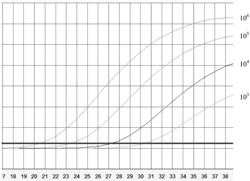
[0070] Embodiment 2: the mensuration of standard curve

[0071] 1. Preparation of Plasmid DNA

[0072] 1.1 Primer design: According to the sequence of HB-1, design primers:

[0073] Upstream primer: 5'-GAGCCTTCTGACCTCACATC-3'

[0074] Downstream primer: 5'-TTGTCCCTGCTCATCCACACC-3'

[0075] Probe sequence: 5'-(FAM)-CCTGAGCCTTCTGACCTCCACA-(TAMRA)-3'.

[0076] 1.2PCR amplification:

[0077] Select the cDNA of the bone marrow specimen with HB-1 gene expression, use this cDNA as a template, and perform PCR amplification in the PCR instrument of Biorad S1000. The amplification conditions are as shown in Table 9:

[0078] Table 9:

[0079] Element content 10×buffer 5μl Upstream and downstream primers (10uM) 1μl each

[0080] 10mMdNTP 0.5μl High Fidelity Taq Enzyme 0.5μl Genomic DNA template of patient specimen as above 1μl DEPC water Make up 50μl

[0081] PCR reaction conditions:

[0082] 95°C for 5min→(95°C f...

Embodiment 3

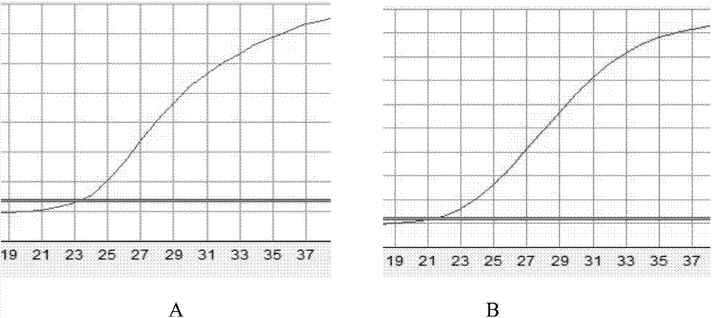
[0130] 1. Sample processing: RNA was extracted from samples 1 and 2 using the Trizol method;

[0131] 2. Preparation of amplification reagents:

[0132] Take out tubes 1-15 from the kit, melt at room temperature and shake to mix, then briefly centrifuge for 1-8 seconds;

[0133] Preparation of sample PCR master mix system:

[0134] System 1: Take 8ul of HB-1 PCR reaction solution, add 12ul of mixed enzyme solution; the number of PCR tubes should be the number of samples;

[0135] System 2: Take 8ul of internal reference PCR reaction solution, add 2ul of mixed enzyme solution I; the number of PCR tubes should be the number of samples;

[0136] System 3: Take 15ul each of HB-1 reference substance 1~4, and add 2ul of enzyme solution II respectively; the number of PCR tubes should be the sum of reference substance 1~4 plus the two controls;

[0137] System 4: Take 15ul each of ABL reference substances 1-4, and add 2ul of enzyme solution II respectively; the number of PCR tubes ...


PUM


Abstract

The invention relates to the field of diagnosis kits, and particularly relates to a pair of primer and kit for detecting the specific gene HB-1 of acute B lymphocyte leukemia. The primers for detecting the HB-1 and a probe are an upstream primer sequence as shown in SEQ ID NO:1, a downstream primer sequence as shown in SEQ ID NO:2 and a probe as shown in SEQ ID NO:3; the 5'end of a nucleotide sequence as shown in the SEQ ID NO:3 has a Fam mark, and the 3'end of the nucleotide sequence has a Tamra mark. The kit disclosed by the invention optimizes the probe sequence, simplifies the experimental step, realizes the single step of a detection method, enhances the experimental stability and realizes the quantitative detection of the gene HB-1.

Description

technical field [0001] The invention relates to the field of diagnostic kits, in particular to a primer and a kit for detecting the specific gene HB-1 of acute B lymphocytic leukemia. Background technique [0002] HB-1 gene is a newly discovered minor histocompatibility antigen gene. The HB-1 gene is located in the 5q32 region of chromosome 5, with a total length of about 8.5kb, including 2 exons, and a transcription product of 382bp, encoding a protein product with 58 amino acid residues. The coding of HB-1 antigenic peptide is initiated by CUG codon, which translates a short peptide protein containing 41 amino acids. So far, literature reports have only found that HB-1 is expressed on the surface of leukemia cells of acute B lymphoblastic leukemia and B cells transformed by Epstein-Barr virus, and is not expressed in normal B cells, nor in T cells, monocytes, and fibroblasts. . [0003] In 1999, Dolstra and other scholars used real-time fluorescence quantitative PCR to ...

Claims


Application Information

Patent Timeline
Patent Type & Authority Applications(China)
IPC IPC(8): C12Q1/68C12N15/11
CPCC12Q1/6886C12Q2600/118C12Q2600/158
Inventor 任汉云王清云李渊邱志祥戢莉刘微梁赜隐
Owner PEKING UNIV FIRST HOSPITAL
Why Eureka
Try Eureka