Dynamic PET image denoising method based on combined image guiding

A technology for combining images and images, applied in image enhancement, image data processing, instruments, etc., can solve the problems of inability to obtain PET image quality, and achieve the effects of assisting clinical diagnosis, reducing noise, and improving quality

Inactive Publication Date: 2014-07-30
SOUTHERN MEDICAL UNIVERSITY
View PDF2 Cites 22 Cited by
  • Summary
  • Abstract
  • Description
  • Claims
  • Application Information

AI Technical Summary

Problems solved by technology

Related methods in the prior art s...

Method used


Image

  • Dynamic PET image denoising method based on combined image guiding
  • Dynamic PET image denoising method based on combined image guiding
  • Dynamic PET image denoising method based on combined image guiding

Examples


Embodiment 1

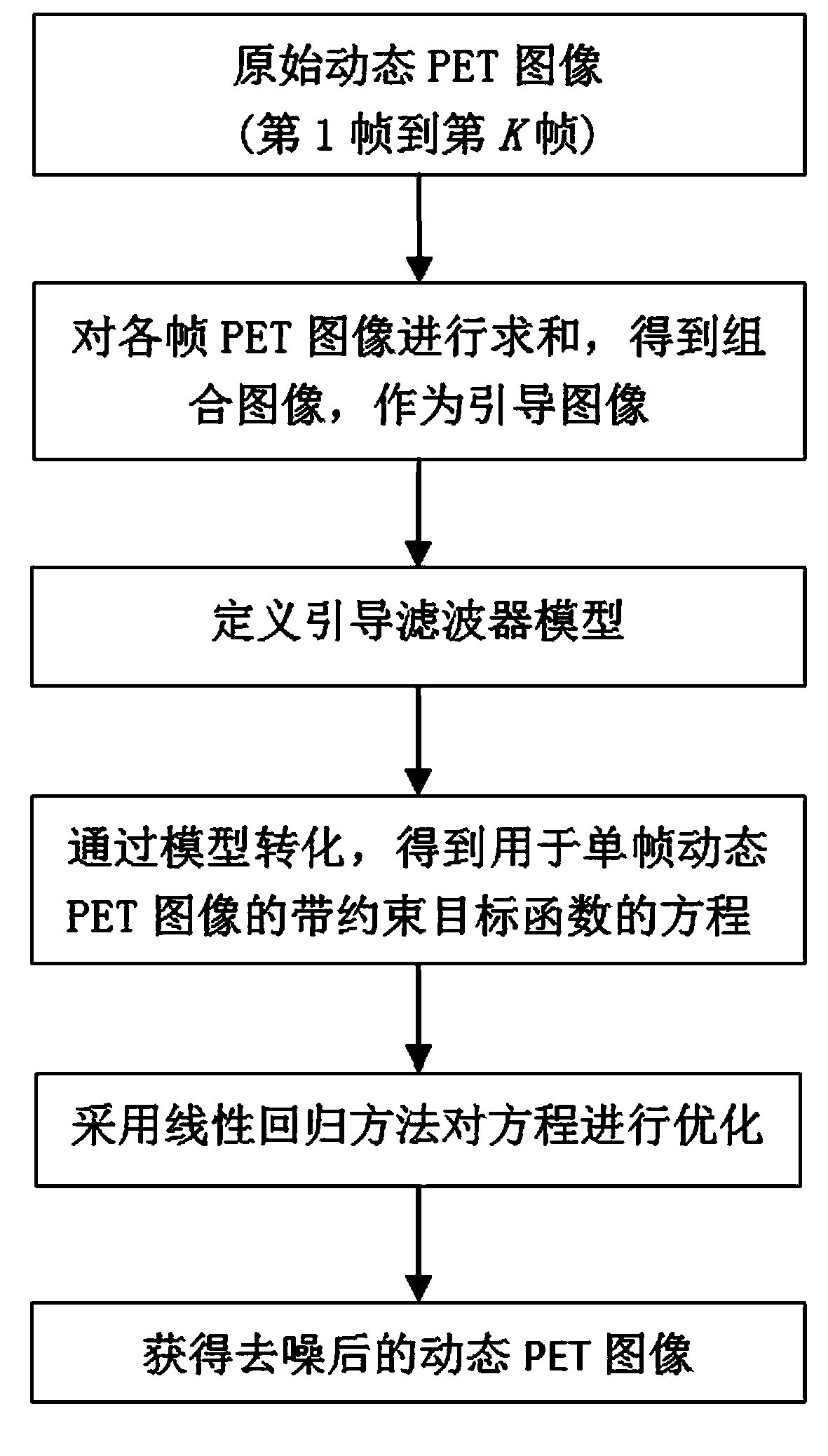
[0040] A dynamic PET image denoising method based on combined image guidance, such as figure 1 shown, including the following steps.

[0041] (1) Use the PET imaging equipment to perform dynamic scanning and reconstruct the dynamic PET image. The dynamic PET image consists of each frame image in the sequence (the first frame to the first frame K frame) composition. The experimental data collection method of the present invention is full three-dimensional collection.

[0042] (2) According to the dynamic PET image acquired in step (1), the combined image is calculated.

[0043] Step (2) is specifically to obtain a combined image by accumulating and summing the pixel values ​​of corresponding pixels in each frame of the PET image in the dynamic sequence.

[0044] (3) Build a guided filter model.

[0045] The guided filter model of the component in step (3) is a linear model, specifically assuming that the filter output is the boot image at the center of the pixel the w...

Embodiment 2

[0062] Experiments are carried out using the method of the present invention and the method in the prior art, and relevant comparison results are obtained.

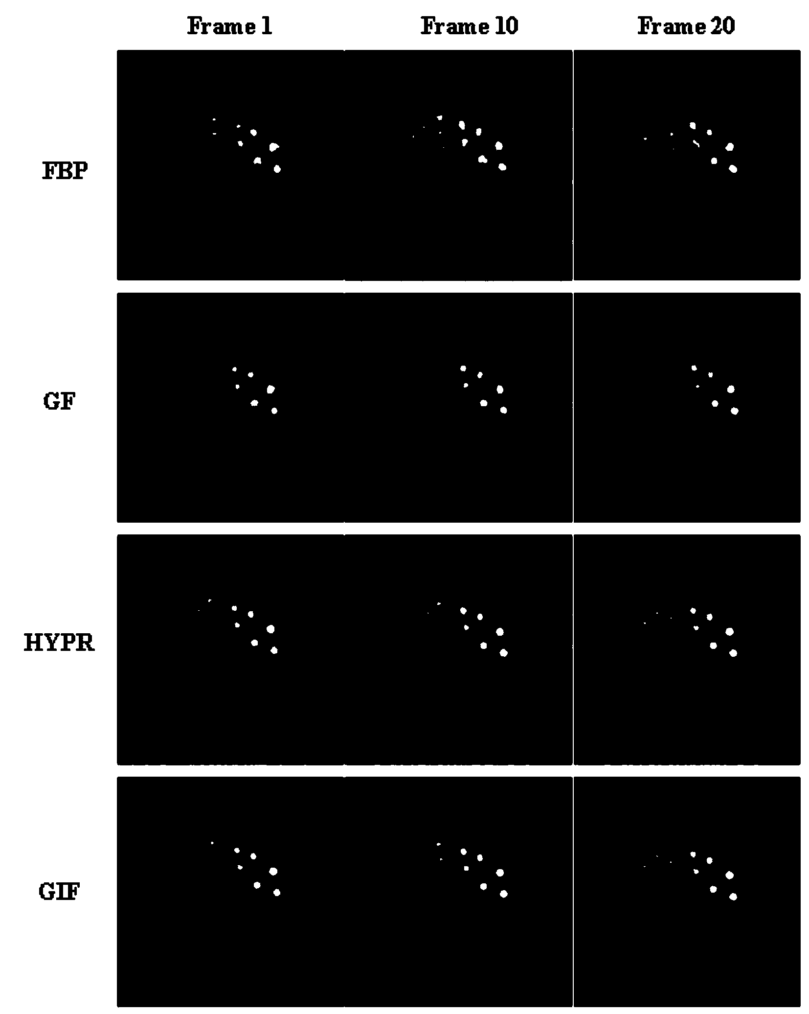
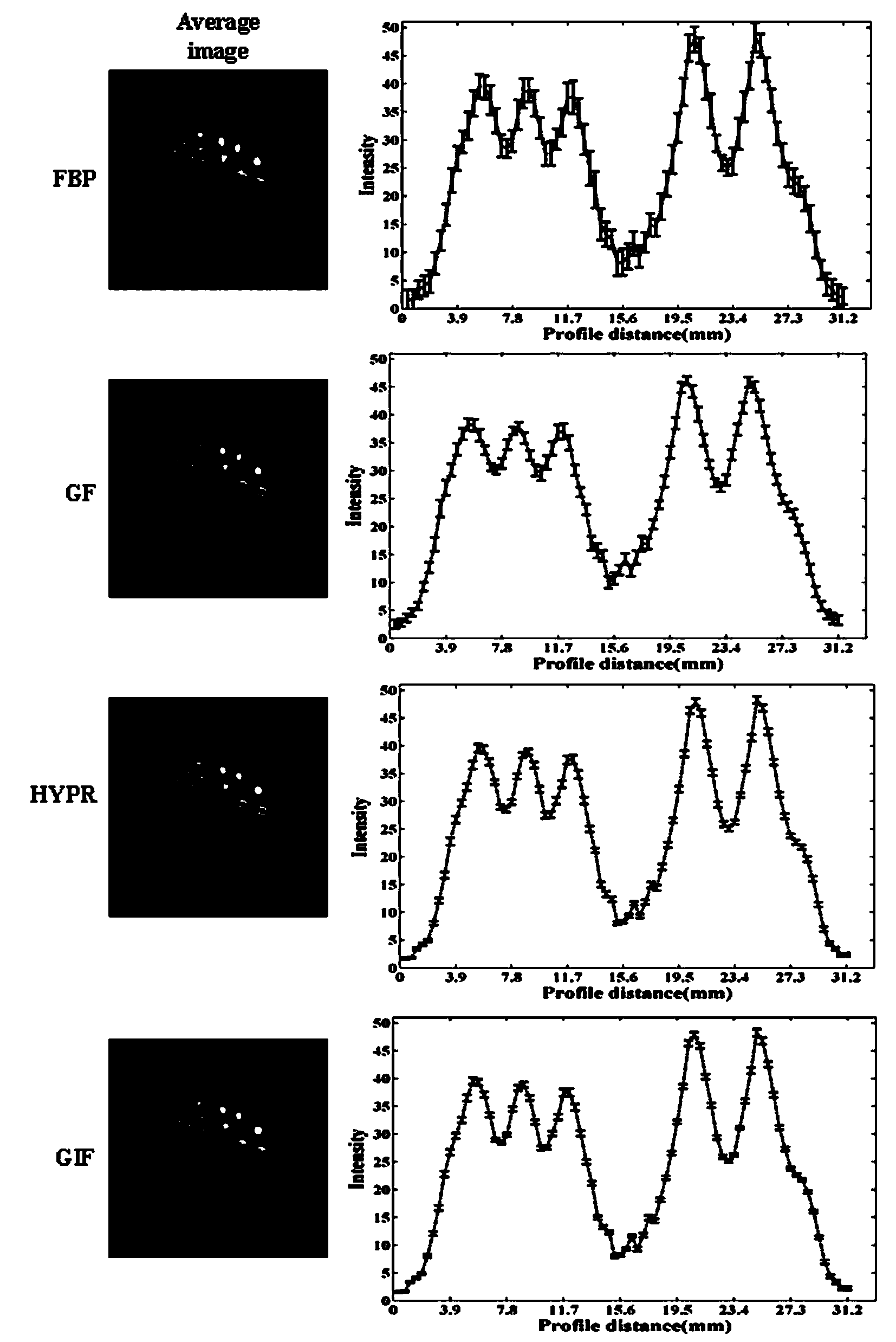
[0063] figure 2 The reconstructed noised and denoised images of scanned Derenzo phantoms are shown. The Derenzo phantom is machined in acrylic and has a cylindrical diameter of 40mm and a length of 13mm. There are multiple circular holes with different diameters in the phantom (diameters are 0.8, 1.0, 1.25, 1.5, 2.0, 2.5mm), and these circular holes are arranged together according to the same diameter. Inject a total activity of 18.54MBq in the Derenzo phantom 18 F-FDG solution, and scanned on a small animal InvoenmicroPET at the PET Center of Nanfang Hospital. The PET system was set as the default setting, and the phantom was scanned dynamically for 20 minutes, with a one-minute interval between each frame, for a total of 20 frames. The reconstruction method is filter backprojection (FBP), and the final image data i...

Embodiment 3

[0067] Another experiment was carried out by adopting the method of the present invention and the method in the prior art to obtain relevant comparison results.

[0068] Figure 5 are scanned NEMA NU4-2008 IQ physical phantom reconstructed noisy images and denoised images processed by different methods. This physical phantom is mainly used for quantitative evaluation of different methods. It is made of plexiglass and has a cylindrical diameter of 30mm and a length of 50mm. It contains a cylindrical cavity with a diameter of 30mm and a height of 30mm and a solid body with a height of 20mm. part. The solid part has five fillable cavity rods with diameters of 1, 2, 3, 4 and 5 mm in sequence, and the cavity rods communicate with the cylindrical cavity. Inject 21ml of the active concentration of 174.8kBq / ml into the phantom 18 F-FDG solution, the total activity obtained was 3.67MBq. Scans were performed on small animal InvoenmicroPET at the PET Center of Nanfang Hospital. The ...


PUM


Abstract

The invention discloses a dynamic PET image denoising method based on combined image guiding. The method comprises the first step of utilizing a PET imaging device to carry out dynamic scanning and reestablishing a dynamic PET image, the second step of calculating and obtaining a combined image according to the reestablished PET image obtained in the first step, the third step of defining a guiding filter model and a kernel function of the guiding filter model, the fourth step of regarding the combined image obtained in the second step as a guiding image, and converting the model obtained in the third step to obtain an equation with a constraint target function for the single-frame dynamic PET image, and the fifth step of carrying out calculation to obtain the denoised dynamic PET image based on the linear regression method and overall parameter selection on the equation according to the result obtained in the fourth step. According to the method, the combined image serves as the guiding image, the noise of the single-frame dynamic PET image can be effectively reduced, and the quality of the dynamic PET image can be greatly improved.

Description

technical field [0001] The invention relates to an image denoising method of medical images, in particular to a dynamic PET image denoising method based on combined image guidance. Background technique [0002] Positron Emission Tomography (PET) is one of the most advanced clinical examination techniques in the field of medical imaging, which can diagnose diseases at the molecular level. Dynamic PET imaging can not only show the spatial distribution of radiotracer concentration, but also reveal the dynamic process of tracer metabolism over time. By applying the kinetic model, kinetic parameters with rich meaning can be obtained, thus providing guidance for disease diagnosis, treatment and drug development. However, in dynamic PET imaging, in order to track changes in the tracer activity of tissues and organs in time, the scanning time of each frame of dynamic PET image is extremely short, and the coincidence events of each frame are correspondingly reduced, so that the sing...

Claims


Application Information

Patent Timeline
IPC IPC(8): G06T5/00G06T5/50A61B6/03
Inventor 路利军胡德斌马晓勉辜承慰马建华陈武凡
Owner SOUTHERN MEDICAL UNIVERSITY
Why Eureka
Try Eureka