Immunofluorescence double labeling method based on same species source first antibody
What is AI technical title?
AI technical title is built by PatSnap AI team. It summarizes the technical point description of the patent document.
An immunofluorescence and double labeling technology, applied in the fields of biomedicine and biotechnology, to avoid cross-reaction, avoid immune cross-reaction, and solve the limited source of antibody species
Inactive Publication Date: 2014-08-20
FOURTH MILITARY MEDICAL UNIVERSITY
View PDF0 Cites 9 Cited by
Summary
Abstract
Description
Claims
Application Information
AI Technical Summary
This helps you quickly interpret patents by identifying the three key elements:
Problems solved by technology
Method used
Benefits of technology
Problems solved by technology
[0006] In order to solve the problem that the existing immunofluorescence double labeling technology must use two different kinds of primary antibodies, the present invention provides a new immunofluorescence chemical staining technology, which uses limited antibody species sources and ordinary fluorescent microscopes to realize the same species Double labeling with primary antibodies of genus origin in the same specimen
Method used
Image
Click on the blue label to locate the original text in one second.
Reading with bidirectional positioning of images and text.
Examples
Embodiment l
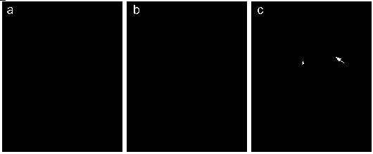
[0034] Embodiment 1: The co-expression of TH (A protein) and P75 (B protein) was detected by the immunofluorescence double-labeling technique described in the present invention, and the primary antibodies used were all rabbit-derived , including the following steps:
[0035] (1) Determine the minimum working concentration of rabbit anti-TH antibody:
[0036] (a) Flat-frozen tissue sections of the coeruleus coeruleus in the midbrain of neonatal mice were washed in 0.01 mol / L PBS three times for 5 minutes each time;
[0037] (b) Add normal goat serum blocking solution and incubate in a humid box at 37°C for 60 minutes. Pay attention to keep the sample moist, and be sure to avoid drying the tissue slices, otherwise a high background will be produced;
[0038] (c) According to the concentration recommended in the instruction manual of rabbit anti-TH antibody (Sigma, Cat. No. T-8700), dilute the antibody in a two-fold gradient with antibody diluent (containing 1% BSA and 0.3% T...
Embodiment 2
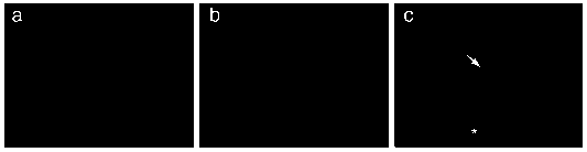
[0055] Example 2: The cultured neural stem cell slides were detected by the immunofluorescence double labeling technique of the present invention to detect the co-expression of Ki67 (A protein) and BrdU (B antigen). The primary antibodies used were all mouse-derived, including the following steps:
[0056] (1) Determine the minimum working concentration of mouse anti-Ki67 antibody:
[0057] (a) The slides of cultured neural stem cells were fixed in 4% paraformaldehyde for 20 minutes, and washed three times with 0.01 mol / L PBS for 5 minutes each time;
[0058] (b) Add normal goat serum blocking solution and incubate in a humid box at 37°C for 30 minutes;
[0059] (c) According to the recommended concentration of the mouse anti-Ki67 antibody (Abcam, Cat. No. ab6526), use the antibody diluent to dilute the antibody in a two-fold gradient. The specific dilution ratios are 1:200, 1:400, 1:800, 1:1500, 1:3000, add to the cell slides to be tested, and incubate at 4°C for 24 hour...
PUM
Abstract
The invention discloses an immunofluorescence double labeling method based on the same species source first antibody. The method comprises the steps of: (1) conducting gradient dilution of anti A antibody, and determining anti A proteinantibody and measuring the minimum working concentration of the A protein to be detected by using an indirect immunofluorescencestaining method; (2) developing the A protein by using the concentration and an SABC method (using luorescein to labelstreptavidin); (3) applying anti B protein antibody, and developing the B protein by an indirect immunofluorescencestaining method; and (4) conducting microscopic examination by a fluorescencemicroscope, taking photos, overlaying the microscopic examination photos of the A protein and B protein to be detected, and analyzing the expression of the two proteins. The method is simple and easy for operation, realizes high research requirements with low experiment cost, and provides a wide range of applications for the chemical study of clinical immunofluorescence tissue.
Description
technical field [0001] The invention belongs to the application fields of biomedicine and biotechnology, and specifically relates to a method for immunofluorescent double labeling by using two primary antibodies from the same species. Background technique [0002] In biological and medical research, immunofluorescent chemical staining is one of the most commonly used technical means. Its principle is to use the principle of specific binding of antigen and antibody, combine with fluorescein labeled on the antibody, and irradiate the fluorescein with excited light. And it emits bright fluorescence (yellow-green or orange-red) so that the localization, qualitative and quantitative information of tissue, intracellular polypeptide or protein antigen can be visualized under a fluorescent microscope. [0003] During the research process, it is often necessary to detect two antigens / proteins (such as protein A and protein B) simultaneously in the same cell or tissue sample to clarif...
Claims
Application Information
Patent Timeline
Application Date:The date an application was filed.
Publication Date:The date a patent or application was officially published.
First Publication Date:The earliest publication date of a patent with the same application number.
Issue Date:Publication date of the patent grant document.
PCT Entry Date:The Entry date of PCT National Phase.
Estimated Expiry Date:The statutory expiry date of a patent right according to the Patent Law, and it is the longest term of protection that the patent right can achieve without the termination of the patent right due to other reasons(Term extension factor has been taken into account ).
Invalid Date:Actual expiry date is based on effective date or publication date of legal transaction data of invalid patent.

