Eureka AIR delivers breakthrough ideas for toughest innovation challenges, trusted by R&D personnel around the world.

Kit for detecting mycobacterium tuberculosis infection and monitoring clinical treatment effect and application of kit

A technology for detection of Mycobacterium tuberculosis and infection, applied in the field of biomedical testing, can solve the problem of the decline in the number of tuberculosis antigen-specific immune cells

Active Publication Date: 2014-09-03
SHANGHAI JIAOTONG UNIV SCHOOL OF MEDICINE
View PDF4 Cites 17 Cited by
  • Summary
  • Abstract
  • Description
  • Claims
  • Application Information

AI Technical Summary

Problems solved by technology

[0005] In the course of clinical anti-tuberculosis treatment, the load of Mycobacterium tuberculosis in the body of tuberculosis patients decreases with the progress of effective treatment, which leads to a decrease in the number of tuberculosis antigen-specific immune cells in the body

Method used


Image

  • Kit for detecting mycobacterium tuberculosis infection and monitoring clinical treatment effect and application of kit
  • Kit for detecting mycobacterium tuberculosis infection and monitoring clinical treatment effect and application of kit
  • Kit for detecting mycobacterium tuberculosis infection and monitoring clinical treatment effect and application of kit

Examples


Embodiment 1

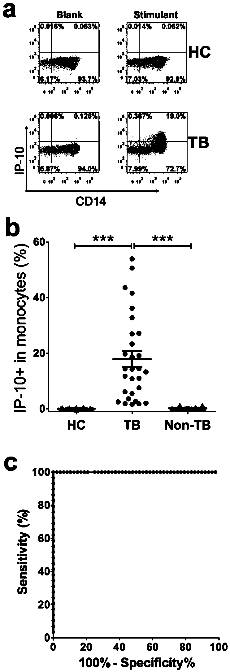
[0049] Example 1: Detection of antigen-specific IP-10 by intracellular cytokine staining.

[0050] The experimental steps are as follows:

[0051] 1. The peripheral blood of patients with active tuberculosis was collected in anticoagulant tubes, and separated by Ficoll density gradient centrifugation to obtain PBMCs.

[0052] 2. PBMCs were washed twice with 5-10ml PBS or RPMI1640, resuspended in RPMI1640 containing 10% FBS, counted the number of cells, and adjusted to a final concentration of 2.5×10 6 / ml.

[0053] 3. Add 100 μl of cell suspension to each well of a 96-well cell culture plate, that is, 0.25×10 6 cell. Mycobacterium tuberculosis-specific antigens or polypeptides were added to the experimental wells, the final concentration of the antigen or single polypeptide was 10 μg / ml, and the final concentration of each polypeptide in the polypeptide library was 2 μg / ml. Add 2.5 μg / ml PHA to the positive control wells, and add the same volume of RPMI1640 medium containi...

Embodiment 2

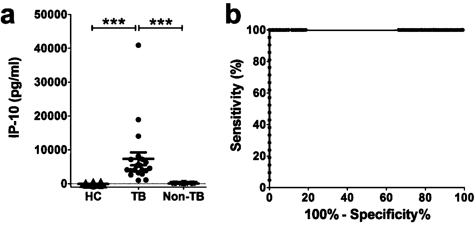
[0064] Example 2: Detection of antigen-specific IP-10 by ELISA.

[0065] The experimental steps are as follows:

[0066] 1. The peripheral blood of patients with active tuberculosis, patients with latent tuberculosis infection, normal people and non-tuberculosis pulmonary disease controls were collected in anticoagulant tubes, and separated by Ficoll density gradient centrifugation to obtain PBMCs.

[0067] 2. PBMCs were washed twice with 5-10ml PBS or RPMI1640, then resuspended in RPMI1640 containing 10% FBS, counted the number of cells, and adjusted to a final concentration of 2.5×10 6 / ml.

[0068] 3. Add 100 μl of cell suspension to each well of a 96-well cell culture plate, that is, 0.25×10 6 PBMC cells. Mycobacterium tuberculosis-specific antigens or polypeptides were added to the experimental wells, the final concentration of the antigen or single polypeptide was 10 μg / ml, and the final concentration of each polypeptide in the polypeptide library was 2 μg / ml. Add 2....


PUM


Abstract

The invention discloses a kit for detecting the mycobacterium tuberculosis infection and monitoring a clinical treatment effect. The kit provided by the invention comprises a specific antibody, namely an IP-10 antibody and / or CD14 antibody, an antigen irritant and a positive contrast irritant. The kit disclosed by the invention can be used for diagnosing an active tuberculosis patient or a tuberculosis latent infection patient and is not affected by BCG (bacillus calmette-guerin) inoculation. The sensitivity and the specificity of the kit for diagnosing the active tuberculosis patient are higher than those of a commercial T-SPOT.TB kit and can be up to 100 percent. After the antituberculosis therapy is performed for 1 month, the detection rates of over 80 percent of clinical tuberculosis patients are converted to be negative, so that the kit can be used for detecting the clinical antituberculosis treatment effect.

Description

technical field [0001] The invention belongs to the field of biomedical testing, and in particular relates to a kit for detecting mycobacterium tuberculosis infection and monitoring clinical treatment effects. Background technique [0002] Tuberculosis is mainly caused by Mycobacterium tuberculosis infection. In 2013, the World Health Organization (WHO) reported that there were about 12 million tuberculosis patients worldwide, of which 8.6 million new tuberculosis cases and 1.3 million deaths occurred in 2012. Tuberculosis poses a serious threat to human health. my country is one of the five high-burden countries of tuberculosis, and tuberculosis seriously hinders my country's economic and social development. [0003] One of the difficulties in TB prevention and control is the lack of rapid and accurate diagnostic methods. Traditional tuberculosis diagnosis methods include tuberculin skin test (TST), acid-fast staining of tuberculosis sputum smear, tuberculosis sputum cult...

Claims


Application Information

Patent Timeline
Patent Type & Authority Applications(China)
IPC IPC(8): G01N33/68
CPCG01N33/5695G01N2800/52
Inventor 肖洋炯王颖沈浩
Owner SHANGHAI JIAOTONG UNIV SCHOOL OF MEDICINE
Why Eureka
Eureka Blog
Learn More