Cellular analysis of body fluids

A technology of cells and body fluids, applied in the direction of individual particle analysis, material analysis, material excitation analysis, etc., which can solve problems that are not common and difficult to analyze

Inactive Publication Date: 2014-09-03
ABBOTT LAB INC
View PDF3 Cites 4 Cited by
  • Summary
  • Abstract
  • Description
  • Claims
  • Application Information

AI Technical Summary

Problems solved by technology

Analysis of samples with significantly lower concentrations of cells, whether by flow cytometry or other techniques, is more difficult and therefore less common

Method used


Image

  • Cellular analysis of body fluids
  • Cellular analysis of body fluids
  • Cellular analysis of body fluids

Examples


Embodiment 1

[0107] Analyzing Low Cell Count Samples

[0108] A prototype analyzer with a 1:35 (blood:reagent) dilution ratio, 2.3 microliter / second injection rate, and 32 second sample measurement (data collection) resulted in over 20 events collecting a sample of 10 cells / microliter.

[0109] Figure 2A and 2B A scatterplot showing cellular analysis of a bodily fluid sample. Cells were spotted after analysis: WBC = 89 / μL (ref = 81 / μL); RBC = 2012 / μL (ref = 1733 / μL). Figure 3A and 3B Scatterplot showing cellular analysis of another body fluid sample. Cells were spotted after analysis: WBC = 0.95 / μL (ref = 0.33 / μL); RBC = 142 / μL (ref = 122 / μL).

Embodiment 2

[0111] Analysis of Very Low Cell Count Samples

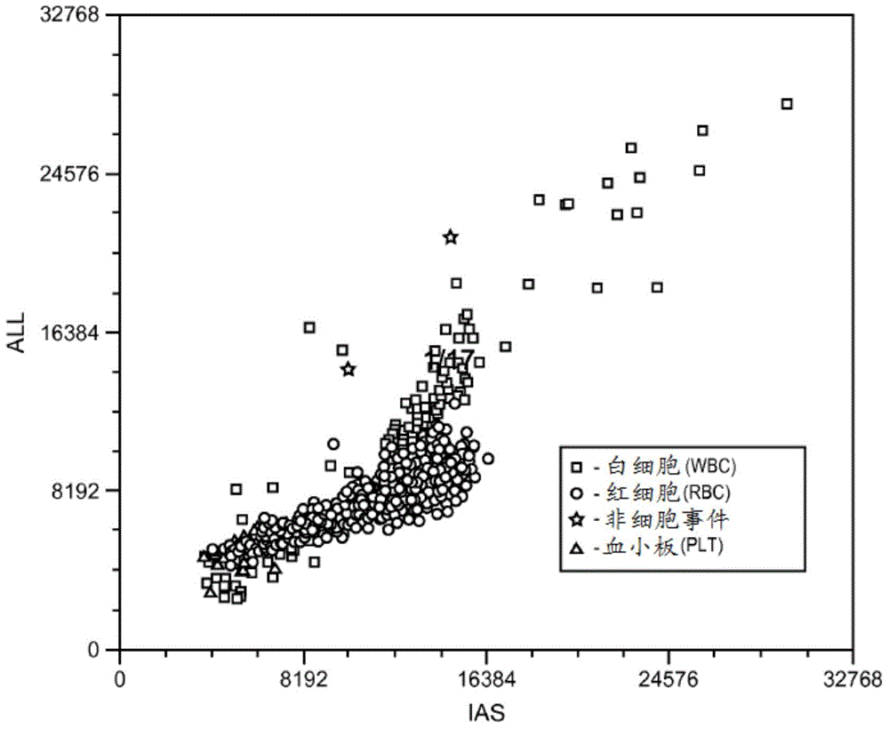
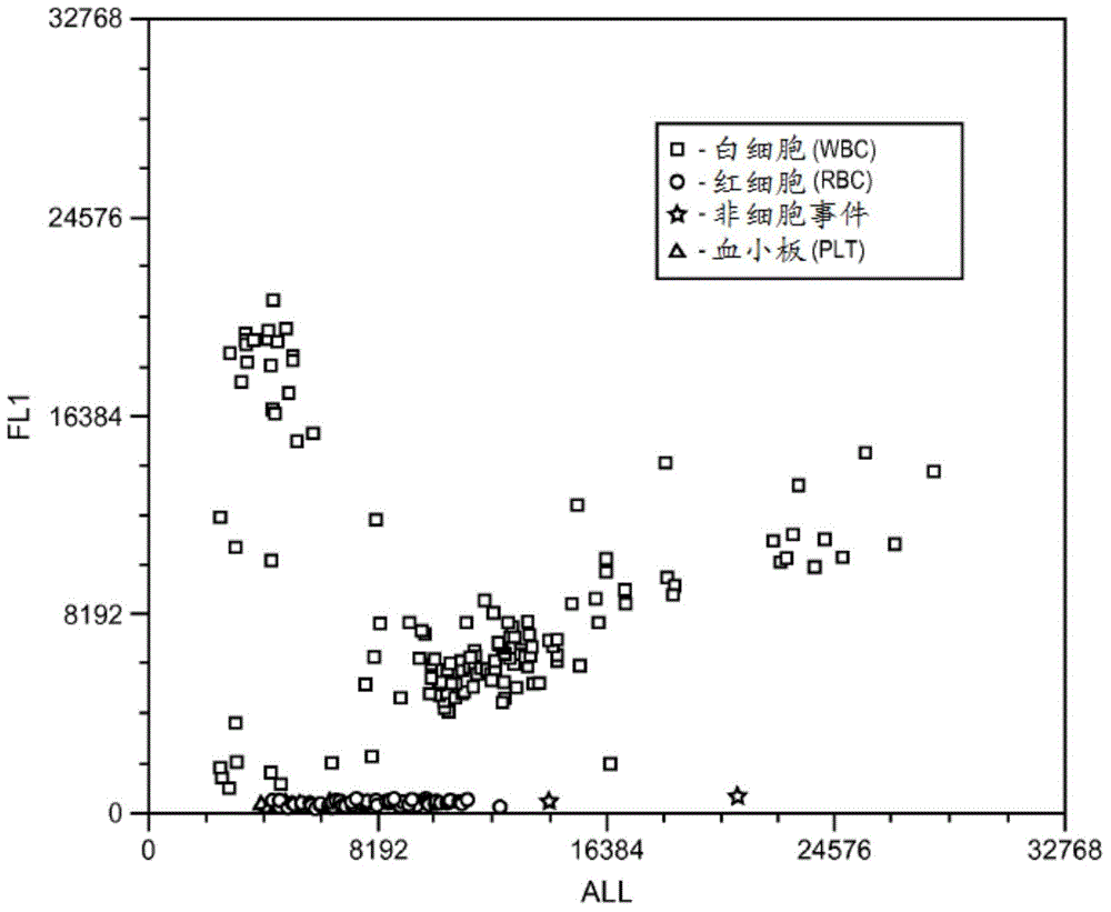
[0112] The limit of detection was further validated in studies using diluted buffy coat samples. Serial dilutions were used to prepare six levels of diluted buffy coat samples. The FL1 versus IAS scatterplots for six horizontally diluted buffy coat samples are shown in Figures 4A-4F . Figure 5 Demonstration of correlation of WBC (measured vs calculated). A good correlation was obtained: (A) for all six levels, Y=1.0239X-4.9 (R 2 =0.9984), and (B) for the three levels with the lowest cell concentration, Y=1.0787X-1.5(R 2 = 0.9598).

Embodiment 3

[0114] Cell Analysis of Body Fluid Samples at 1:35 Dilution Ratio

[0115] A comprehensive body fluid study was performed to evaluate the methods presented herein. A total of 91 body fluid samples, including CSF fluid, pleural fluid, peritoneal fluid, and ascitic fluid, were measured on the prototype analyzer at a 1:35 (blood:reagent) dilution ratio, 2.3 μl / sec infusion rate, and 32-second sample measurement (data collection ) for measurement and analysis. Reference values ​​for WBC and RBC were obtained by manual chamber counting.

[0116] An excellent correlation of WBC was obtained between the method described above and the reference method ( Figure 6 ): (A)Y=1.1305X-9.8411,R 2 = 0.997 (full range, up to approximately 40,000 / μL); (B) Y = 1.017X+7.1395, R 2 =0.9765(2 =0.9663(2 =0.8459 ( Figure 7 ): (A)Y=0.8996X+403.62,R 2 = 0.9595 (full range, up to approximately 200,000 / μL); (B) Y = 1.0833X-11.427, R 2 =0.9513(2 =0.8284(2 =0.6917 (<50 / μL). For RBC analysis, sample...


PUM


Abstract

Herein is provided a method for cellular analysis on hematology analyzers. In various aspects, the methods provide separation and/or differentiation between red blood cells (RBCs) and white blood cells (WBCs) by utilizing a fluorescent dye to selectively stain WBCs such that they emit stronger fluorescence signals. The method provides optimal detection limits on WBCs and RBCs, thereby allowing analysis of samples with sparse cellular concentrations. As few as one reagent may be used to prepare a single dilution for body fluid analysis, in order to simplify the body fluid analysis. Minimal damage to WBCs is attained using the lysis-free approach described in aspects of the disclosure.

Description

[0001] Cross References to Related Applications [0002] This application claims the benefit of priority to US Provisional Patent Application Serial No. 61 / 580,623, filed December 27, 2011; the entire disclosure of which is incorporated herein by reference in its entirety. Background technique [0003] Various methods are used for cell analysis, including visual and / or automated inspection via light or fluorescence microscopy. These types of cellular examination and analysis are typically performed in order to obtain information about the lineage, maturation stage, and cell count of the cells in the sample. [0004] Flow cytometry is a method for identifying and differentiating between different cell types in heterogeneous samples. In a flow cytometer, cells pass through a sensing region one at a time, or nearly one at a time, where each cell is illuminated by an energy source. Typically, a single wavelength light source (eg, laser, etc.) is used as the energy source and one...

Claims


Application Information

Patent Timeline
IPC IPC(8): C12Q1/68G01N33/48C12M3/00G01N1/10
CPCG01N33/5094C12Q1/04G01N15/14G01N21/17G01N21/64G01N33/49G01N2015/0065G01N2015/1006
Inventor 吴炯艾米丽·H.·林杨金平
Owner ABBOTT LAB INC
Why Eureka
Try Eureka